mail_outline sales@mediastorehouse.com
Aortic aneurysm, illustrationAortic aneurysm. Computer illustration showing an aneurysm in the ascending aorta
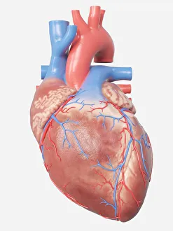
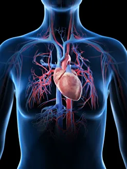
Illustration of the human heart anatomy
Male heart, illustrationIllustration of male heart and arteries
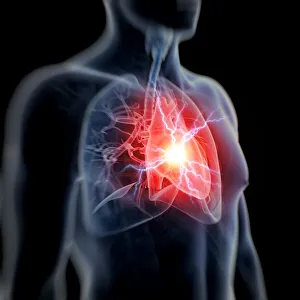
Human heart attack, illustrationHuman heart attack, computer illustration
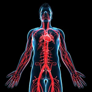
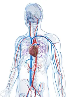
Human vascular system, artworkHuman vascular system, computer artwork
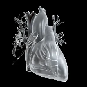
Illustration of glass heart
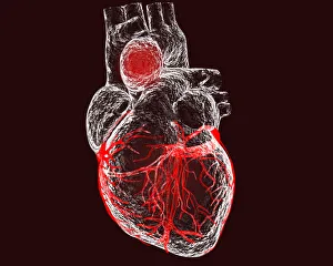
Illustration of the human heart
Female vascular system, illustrationFemale vascular system, computer illustration
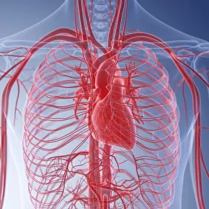
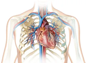
Human heart and chest, illustrationHuman heart with vessels, bronchial tree and cut rib cage. On white background
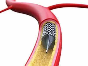
Stent inside of an artery, illustration
Peas in a pod with skeletons, X-ray
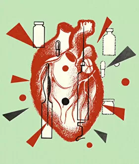
Human Heart and Medicine Containers
One Hand
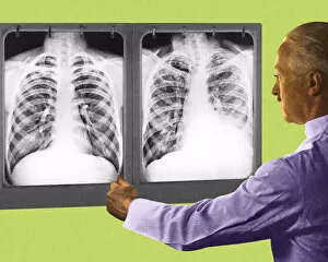
Man Looking at Two Chest X-Rays
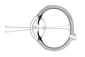
Eye
Cell Biology
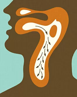
Diagram of Swallowing
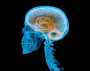
Human skull with brain, illustrationHuman skull with brain. X-ray effect. Side view on black background
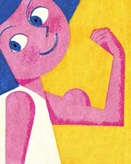
Flexing Muscle
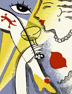
Woman Drinking Champagne
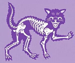
Cat Skeleton
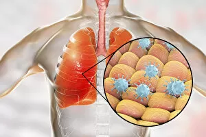
Viral pneumonia, conceptual illustration. Human lungs and close-up view of viruses, the causative agents of pneumonia. Pneumonia can be caused by many types of viruses, including Influenza, MERS
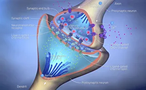
Nerve synapse, illustration3d illustration of a synapse
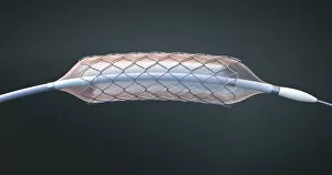
Stent and balloon catheter, illustration
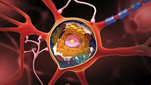
Nerve cell, illustrationNerve cell, cut-away illustration
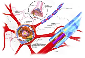
Nerve cell anatomy, illustrationCross-section of a neuron, illustration
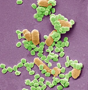
Virus particles and bacteria, SEMVaccinia virus particles and bacteria. Coloured scanning electron micrograph (SEM) of vaccinia virus particles (green). Unlike most viruses, vaccinia replicates in the cells cytoplasm
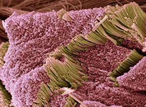
Tendon, SEMTendon, coloured scanning electron micrograph (SEM), showing bundles of collagen fibres. The parallel alignment of the fibres make tendons inelastic but flexible. Tendons attach muscle to bone
Osteocyte, SEMOsteoblast bone cell. Coloured scanning electron micrograph (SEM) of an osteoblast bone cell. Osteocytes are osteoblasts (bone-producing cells) that have become trapped within bone cavities (lacunae)
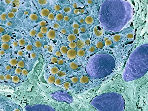
Pancreas tissue, colored scanning electron micrographPancreas tissue. Coloured scanning electron micrograph (SEM) of fractured pancreas tissue. Seen here are zymogen granules (yellow) and cell nuclei (purple)
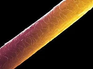
Human hair, semHuman hair (Caucasian, brunette), coloured scanning electron micrograph (SEM). The outer layer of hair (the cuticle) has overlapping scales of keratin
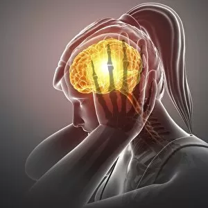
Woman with headache, illustrationWoman with headache, computer illustration
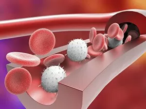
Human artery with blood cells, artworkHuman artery, cut-away computer artwork, showing the artery wall and red and white blood cells
Red blood cells and platelets, SEMRed blood cells and platelets. Coloured scanning electron micrograph (SEM) of human erythrocytes (red blood cells) and a platelet aggregate (orange)
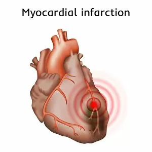
Heart attack, illustrationHeart attack (myocardial infarction), illustration
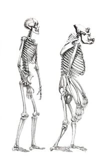
Human skeleton and a skeleton of a monkey, anatomical illustration
Exuvia, remaining larval skin, cast skin of a dragonfly, old skin after moulting
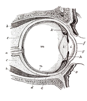
Eye, anatomical illustration
Eye, schematic representation
Acupuncture figure as a shadow