mail_outline sales@mediastorehouse.com
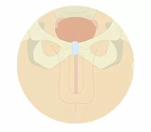
Cross section biomedical illustration of human male reproductive system and pelvis
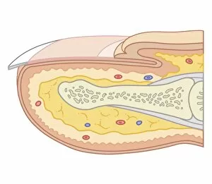
Cross section biomedical illustration of fingernail
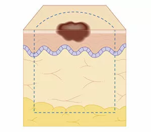
Cross section biomedical illustration of site of skin biopsy
Exuvia, remaining larval skin, cast skin of a dragonfly, old skin after moulting
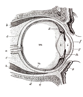
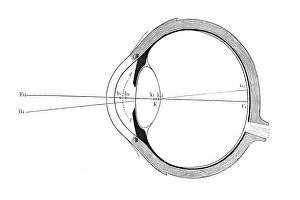
Eye, anatomical illustration
Eye, schematic representation
Wood structure
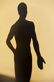
Acupuncture figure as a shadow
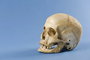
Human skull
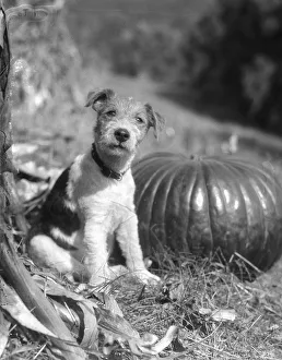
Wire Hair TerrierUNITED STATES - CIRCA 1930s: Portrait Of A Dog, A Wire Hair Terrier Sitting By Pumpkin And Looking At Camera, Making Eye Contact And Looking Cute Outdoors In The Autumn. (Photo by H)
Dolphins jumping out of waterUNITED STATES - CIRCA 1950s: Two dolphins jumping out of water, one with mouth open. (Photo by H. Armstrong Roberts/Retrofile/Getty Images)
Dog holding newspaperUNITED STATES - CIRCA 1950s: Mixed breed dog holding newspaper in mouth. (Photo by H. Armstrong Roberts/Retrofile/Getty Images)
Poodle wearing hat, holding pipe in mouthUNITED STATES - CIRCA 1960s: Poodle dog with pipe in mouth, wearing green paper party hat for St Patricks Day. (Photo by H. Armstrong Roberts/Retrofile/Getty Images)
Terrier wearing hat and glassesUNITED STATES - CIRCA 1950s: Terrier dog wearing spectacles and hat. (Photo by H. Armstrong Roberts/Retrofile/Getty Images)
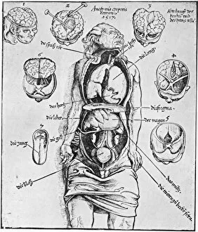
Anatomia Corporis HumaniA diagram of the human brain and other organs. Anatomia Corporis Humani from Spiegl der Arztny by Lorenz Fries, 1518. The diagrams are based on a dissection carried out by German physician Wendelin
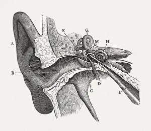
The human ear, wood engraving, published in 1880Anatomy of the human ear: A) auricle, B) External Auditory Canal, C) Tympanic Membrane, D) Tympanic Cavity, E) Malleus, M) Incus, H) Cochlea, G) Semicircular Canals, I) Eustachian Tube
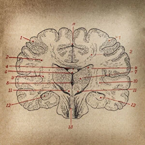
Brain section
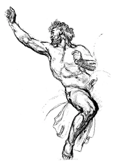
Antique illustration of sketch of Attilas bodyAntique illustration of sketch of Attilas naked body, the character of a Pierre Corneilles drama, 17th century French tragedian
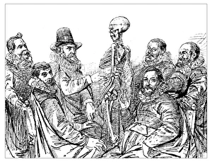
Antique illustration of 17th century anatomy lesson with skeleton: Doctor Egberts teach his students the skeleton structure (from a painting by the 17th century Dutch painter Thomas de Keyser)
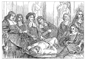
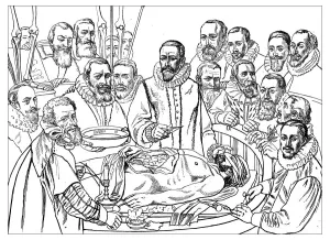
Antique illustration of 17th century anatomy lesson: doctor Professor Ruysch dissects the body of a dead man and his students are around him
Antique illustration of 17th century anatomy lesson: doctor Van Der Meer dissect the body of a dead man and his students are around him at the university of Delft
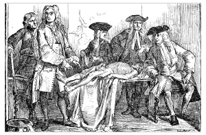
Antique illustration of 18th century anatomy lesson: doctor Professor Roell dissects the body of a dead man and his students are around him