mail_outline sales@mediastorehouse.com
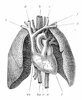
Pulmonary artery engraving 1899Illustrated Natural History of the Three Kingdoms Illustrierte Naturgeschichte der drei Reiche Franz StrAÔé¼ssles - Wilhelm Nitzschke, Stuttgart 1888
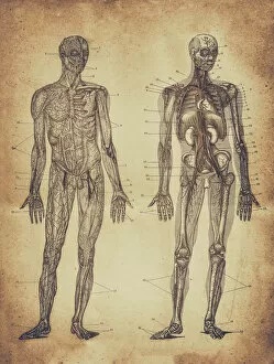
The human skeleton engraving 1895The human skeleton engraving
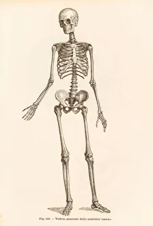
Human skeleton engraving 1899Corso Elementare di Scienze Naturali
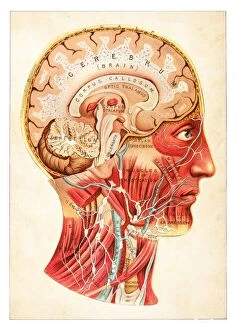
Brain medical illustration 1891The Practical Physician
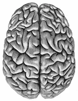
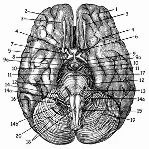
Human brain anatomy engraving 1857Rank, johannes - The human being. 1 - 1894
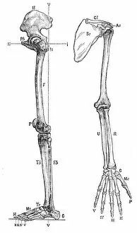
Bones of the arm anatomy engraving 1878Encyclopedia Britannica 9th Edition Vol I New York, Samuel Hall 1878
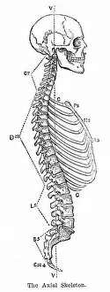
The axial skeleton anatomy engraving 1878Encyclopedia Britannica 9th Edition Vol I New York, Samuel Hall 1878
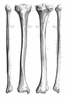
Leg bones anatomy engraving 1866Atlas d anatomie descriptive du corps humain C. Bonamy - Paul Broca Victor Masson et Fils Paris 1866
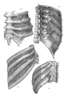
Intercostal nerves anatomy engraving 1866Atlas d anatomie descriptive du corps humain C. Bonamy - Paul Broca Victor Masson et Fils Paris 1866
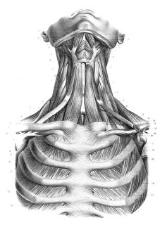
Neck throat anatomy engraving 1866Atlas d anatomie descriptive du corps humain C. Bonamy - Paul Broca Victor Masson et Fils Paris 1866
BrainAntique illustration of a human brain
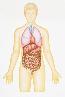
Human male anatomyAntique illustration of a Human male anatomy
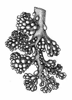
Lung alveoli treeAntique illustration of a Lung alveoli tree
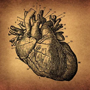
Human Heart EngravingIllustration of a Human Heart Engraving
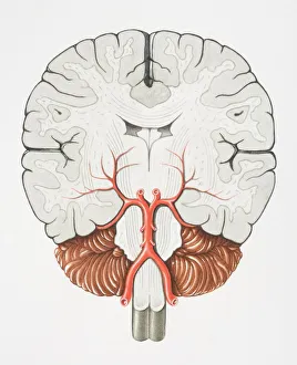
Coronal cross section of the Brain
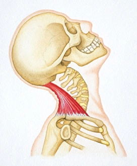
Head bent backwards with bones and relevant neck muscle revealed
Foot flexed with bones and relevant muscle revealed
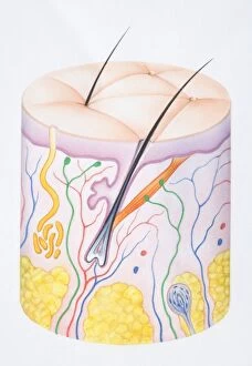
Diagram illustrating the two layers of human skin, epidermis, dermis and hair follicle
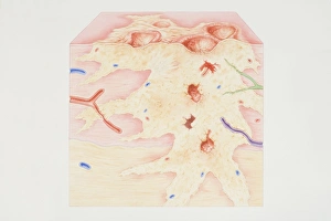
Cross-section diagram of a cancerous tumour including calcium deposits, blood vessels, tumour outgrowth, epithelial layer, ulcerated area, bleeding, nerve fibres, dead tissue and a lymph vessel
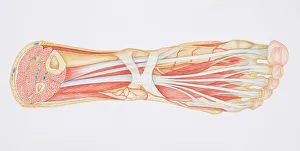
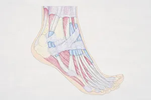
Diagram of the muscles and tendons in the human ankle and foot
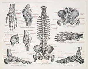
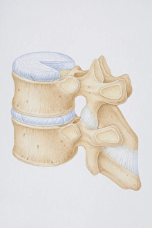
Diagram of a human thoracic vertebrae including the intervertebral disk, vertebral process and ligaments
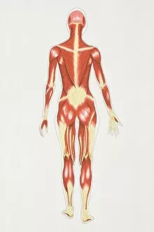
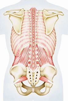
Diagram illustrating muscular structure of female body, rear view
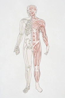
Diagram illustrating the human musculo-skeletal system
Cross-section diagram of human foot, side view
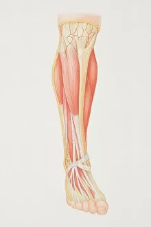
Diagram of lower leg illustrating muscle groups, nerves and veins
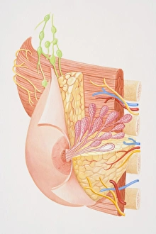
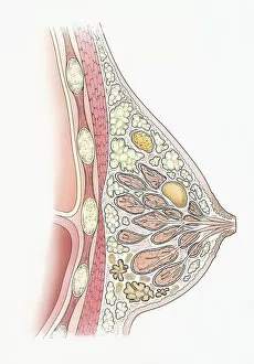
Cross-section diagram of female breast
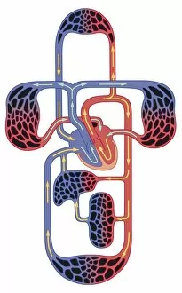
Blood flow diagram
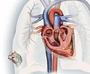
Cross section of human heart and, bottom left, coronary system with aorta, coronary arteries, coronary vein and blood vessels
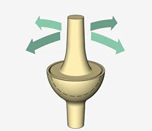
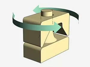
Illustration, ellipsoidal joint, arrows indicating possible directions of movement
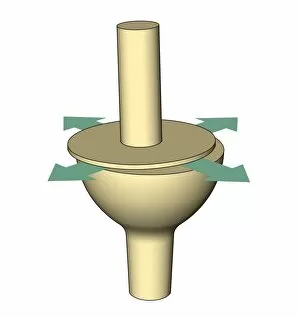
Illustration, pivot joint, arrows indicating possible directions of movement
Illustration, plane joint, arrows indicating possible directions of movement
Human thorax, shoulder girdle, and pelvis posterior view
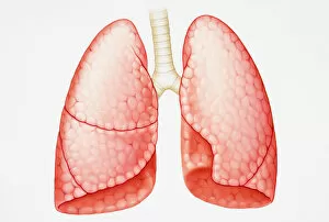
Pair of human lungs
Illustration of human digestive and respiratory systems
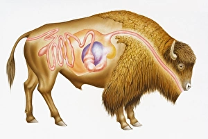
Digitalcross section illustration of European bison (Bison bonasus) showing digestive system
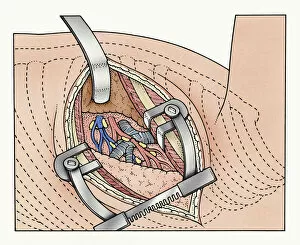
Illustration of abdominal incision between ribs kept open by spreaders exposing human lung and blood vessels
Illustration showing cyst, fibroadenoma, and fibrocystic disease in cross section of human breast
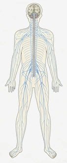
Illustration of human Central Nervous System showing brain, spinal cord, veins and arteries
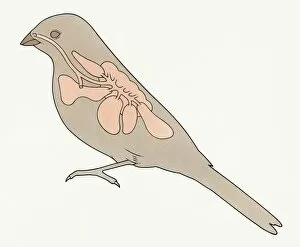
Illustration of respiratory system of bird showing lung, trachea, bronchus, pulmonary alveolus, and syrinx
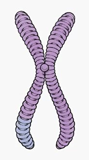
Illustration of human chromosome showing chromatid, centromere, short arm and long arm
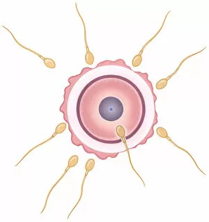
Illustration of human sperm fusing with ovum during conception
Cartoon representing thymus pointing at white blood cells flexing muscles
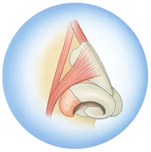
Illustration of septum cartilage inside human nose
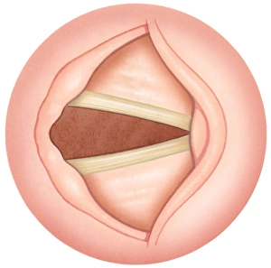
Illustration of open human vocal fold to inhale air, also known as vocal cords
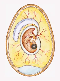
Illustration of yoke sac and amniotic membrane surrounding chicken embryo
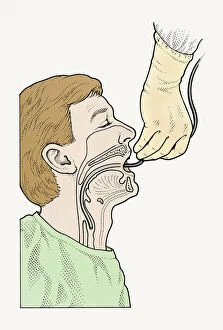
Diagram showing bronchoscope being inserted into patients throat
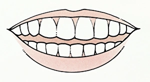
Illustration of mouth showing healthy set of teeth
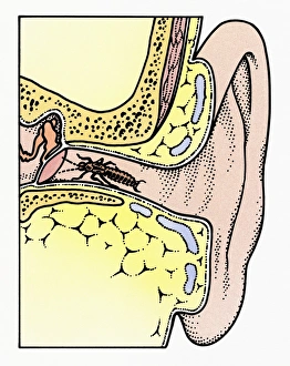
Cross section illustration of Common Earwig (Forficula auricularia) in auditory canal of ear, touching tympanic membrane with antennae