mail_outline sales@mediastorehouse.com
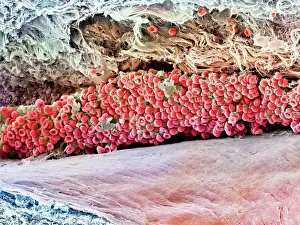
Scanning electron micrograph (SEM) of red blood cellAnatomy, Biology, Blood Clot, Blood Vessel, Cell, Color Image, Fractur, SEM, 85758265
Solar panel with sun
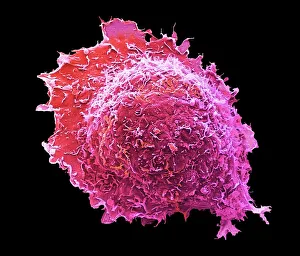
Top of mans head visible behind prison cell window as his hands grip metal bars, front view
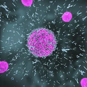
Antibodies attacking virus particles, illustration3d illustration of antibodies attacking virus particles in the bloodstream
Anatomy, Biology, Blood Vessel, Cell, Color Image, Fibrin, Healthcare And MedicineAnatomy, Biology, Blood Vessel, Cell, Color Image, Fibrin, Healthcare, Science Photo Library, 85758208
Osteocyte bone cell, SEMOsteocyte bone cell. Coloured scanning electron micrograph (SEM) of an osteocyte bone cell (blue) surrounded by bone tissue (grey)
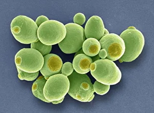
Yeast cells, SEMYeast cells. Coloured scanning electron micrograph (SEM) of cells of brewer s, or baker s, yeast (Saccharomyces cerevisiae). This fungus consists of single vegetative cells
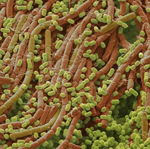
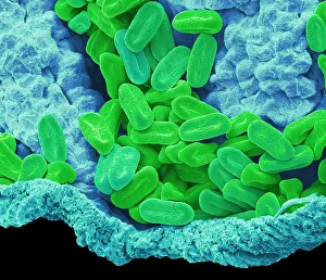
Soil bacteria, SEMSoil bacteria. Coloured scanning electron micrograph (SEM). Bacteria in the soil are directly tied to nutrient recycling especially carbon, nitrogen, phosphorus and sulfur
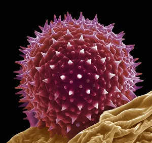
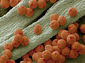
Hollyhock pollen grain, SEMHollyhock pollen grain. Coloured scanning electron micrograph (SEM) of a pollen grain from a hibiscus (Alcea setosa) flower. Pollen grains are the male sex cells of a flowering plant
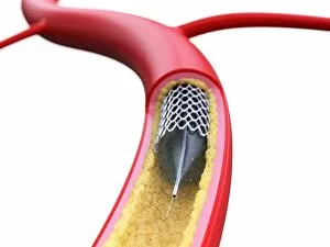
Stent inside of an artery, illustration
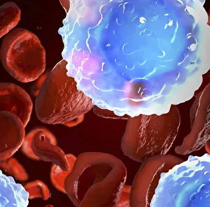
White and red blood cells, illustration3d illustration of white blood cells (leukocytes) in the human body
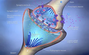
Nerve synapse, illustration3d illustration of a synapse
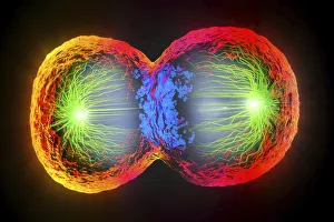
Dividing cell, illustration3d illustration of cell division, cell membrane and a splitting red nucleus
Cell, illustration3d illustration of cell division, cell membrane and a splitting red nucleus
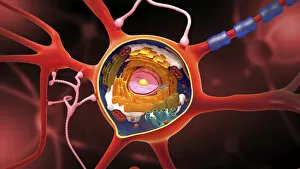
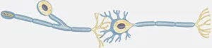
Nerve cell, illustrationNerve cell, cut-away illustration
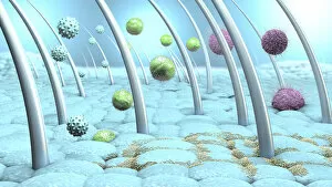
Bacteria and viruses on human skin, illustration
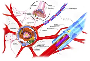
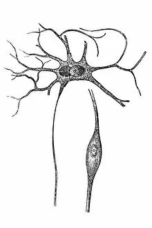
Nerve cell anatomy, illustrationCross-section of a neuron, illustration
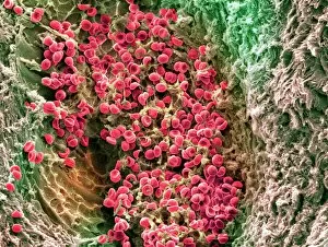
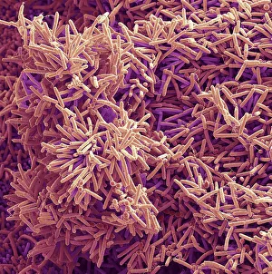
Plaque-forming bacteria, SEMPlaque-forming bacteria, coloured scanning electron micrograph (SEM). Plaque consists of a film of bacteria embedded in a glycoprotein matrix
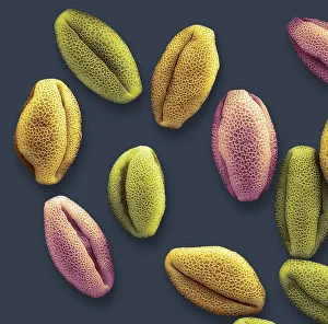
Verbena pollen, SEMVerbena pollen. Coloured scanning electron micrograph (SEM) of pollen grains from verbena bonariensis. Verbena bonariensis is a tall, slender-stemmed perennial
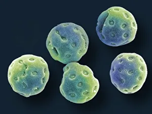
Convolvulus pollen grains, SEMConvolvulus pollen grains. Coloured scanning electron micrograph (SEM) of pollen grains from a convolvulus flower. Convolvulus is a genus of about 200 to 250 species of flowering plants in
Osteocyte, SEMOsteoblast bone cell. Coloured scanning electron micrograph (SEM) of an osteoblast bone cell. Osteocytes are osteoblasts (bone-producing cells) that have become trapped within bone cavities (lacunae)
Water lily pollen grains, SEMWater lily pollen grains. Coloured scanning electron micrograph (SEM) of pollen grains from a water lily flower. Nymphaeaceae is a family of flowering plants, commonly called water lilies
Bellflower pollen, SEMBellflower pollen. Coloured scanning electron micrograph (SEM) of pollen grains from a bellflower (Campanula sp.). Pollen grains are the male gametes (sex cells) of a plant
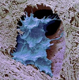
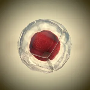
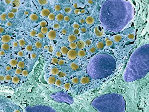
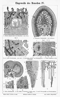
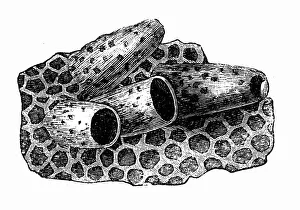
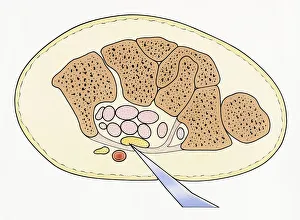
Pancreas tissue, colored scanning electron micrographPancreas tissue. Coloured scanning electron micrograph (SEM) of fractured pancreas tissue. Seen here are zymogen granules (yellow) and cell nuclei (purple)
Cartoon representing thymus pointing at white blood cells flexing muscles
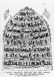
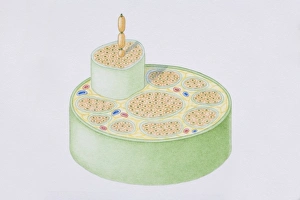
Peoples Beehive1840: The British Beehive, a cartoon depicting the population as members of a behive with the Queen at the top and various other occupations in cells below her
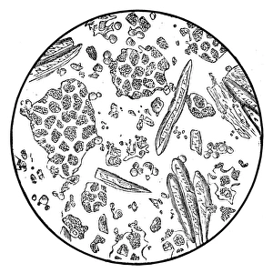
Coffee under microscopeIllustration of a coffee under microscope
Human intestine liver engraving 1895Meyers Konversations-Lexikon. Ein Nachschlagewerk des allgemeinen Wissens, 5th edition 17 volumes Bibliographisches Institut - Leipzig 1895-1897
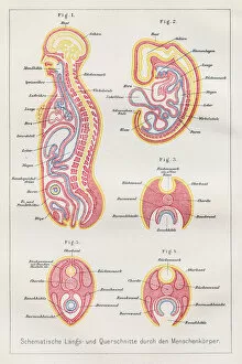
Sections human body anatomy engraving 1857Rank, johannes - The human being. 1 - 1894
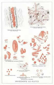
Microscopy of the blood anatomy engraving 1857Rank, johannes - The human being. 1 - 1894
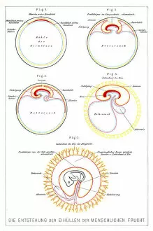
The development of human beings anatomy engraving 1857Rank, johannes - The human being. 1 - 1894
Cell of the queen
Ductal cells refer to cells lining the pancreatic duct
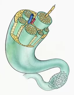
Cross-section diagram of a human nerve fascicle, including a bundle of nerve fibres, blood vessels and myelin sheath
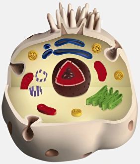
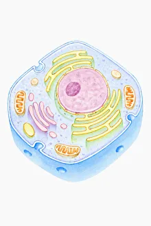
Structure of an animal cell, digital illustration
Cross section illustration of human cell
Illustration of peripheral nerves showing ganglion, nerve Fibers, myelin sheath, Arteries, veins and fat cells
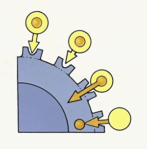
Illustration showing excess Cholesterol in bloodstream over-saturating body cells
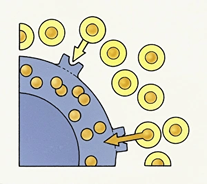
Illustration showing cholesterol molecules passing from blood cells into body cell membrane to regulate blood cholesterol level
Illustration of human nerve cell cross section showing dendrite, soma, axon, nucleus, nodes and myelin sheath
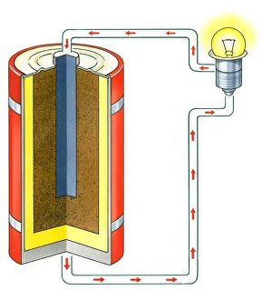
Illustration of showing electrons flowing from negative terminal of dry cell battery to lightbulb
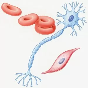
Illustration of human muscle cell, nerve cell, and red blood cells
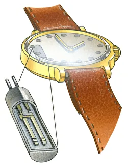
Illustration of solar cell used in light-powered quartz crystal wrist watch
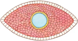
Illustration of cross section showing how early stage embryo of Flatworm (Phylum Platyhelminthes) differentiates into three layers of cell tissue, ectoderm, mesoderm and endoderm
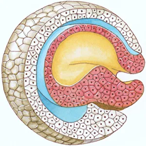
Illustration of cross-section of embryo forming the blastophore
Illustration showing cross section of double layer of dinosaur cell membrane
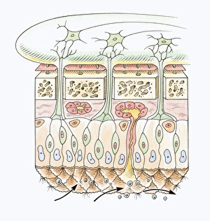
Cross section illustration of human olfactory system
Illustration showing cross section of severed median nerve in wrist caused by sharp piece of glass penetrating skin