mail_outline sales@mediastorehouse.com
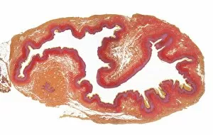
Cervix, LMCervix. Low power lght micrograph (LM) of the cervix. The cervix is the narrow inferior portion of the uterus. The part which projects into the vagina is seen here
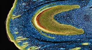
Developing nail, LMDeveloping nail. Light micrograph (LM) of longitudinal section through a fetal finger tip to show the developing nail. The large area of green-yellow nail bed epithelium is tipped by the developing
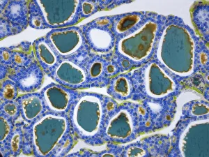
Thyroid, LMThyroid gland. Light micrograph (LM) of a thyroid gland showing the follicles. The follicles are lined by a single layer of cuboidal epithelial cells (blue)
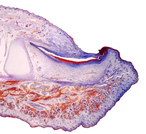
Fingertip, LMFingertip. Light micrograph (LM) of a section through the fingertip. The nail (orange) is at top center, with the nail root below. The nail bed is dark purple and is continous with the epithelium
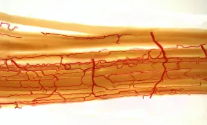
Blood supply to muscles, LMBlood supply to muscles. Light micrograph (LM) showing blood supply to muscle fibers. The muscle fibers (yellow) have been teased apart to reveal the capillary bed (red)
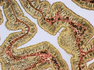
Fallopia tube, LMFallopian tube. Light micrograph (LM).The fallopian tube, or oviduct, conveys the egg from the ovary to the uterus. Ciliated columnar epithelium is yellow
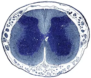
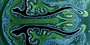
Spinal cord, LMSpinal cord. Light micrograph (LM) of a cross-section through the human spinal cord in the lumbar region. The spinal cord consists of a butterfly-shaped core (dark blue) known as grey matter
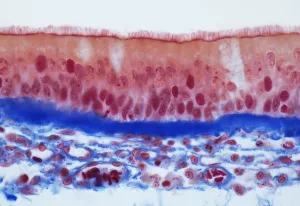
Trachel epithelium, LMTrachea epithelium. Light micrograph (LM) of a vertical section through the pseudostratified columnar epithelium from the trachea
Nasal sinuses, LMNasal sinuses. Light micrograph (LM) of the nasal sinuses ( lined by cyan epithelium ) and the supporting cartilages (green). Bone tissue is identified by the blue bone marrow