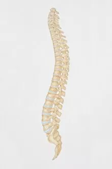
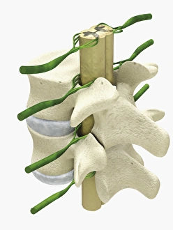
Normal lateral view of the lumbar vertebrae showing spinal nerve roots
Detail, Anatomy, Diagram, Illustration, Spine, Bone, Medical, Biology, Disk, Normal, Cut Out, White Background, Vertical, Artwork, Color Image, Close Up, Healthcare And Medicine, The Human Body
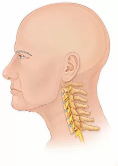
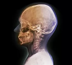
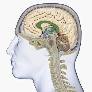
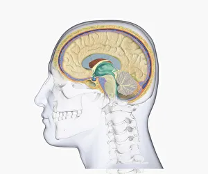
Normal lateral view of a mans head and neck with a skull
Anatomy, Diagram, Illustration, Spine, Medical, Neck, Biology, Disks, Disk, See Through, Cut Out, White Background, Profile, Vertical, Artwork, Male Likeness, Color Image, Healthcare And Medicine
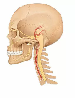
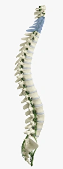
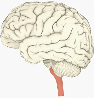
Normal side view of an adult skull showing the spinal cord
Anatomy, Diagram, Illustration, Spine, Bone, Teeth, Skull, Medical, Biology, Disks, Side View, Cut Out, White Background, Vertical, Artwork, Color Image, Human Skull, Healthcare And Medicine
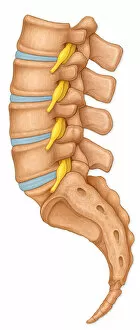
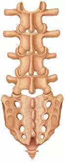
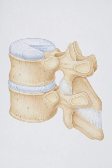
Normal posterior view of the lumbar spine and sacrum
Detail, Anatomy, Diagram, Illustration, Spine, Bone, Medical, Facets, Bones, Biology, Rear View, Cut Out, White Background, Vertical, Artwork, Color Image, Close Up, Healthcare And Medicine
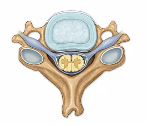
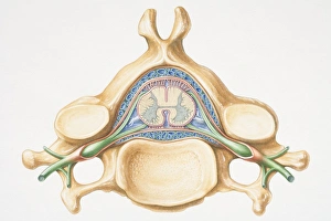
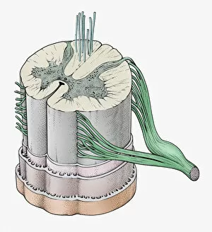
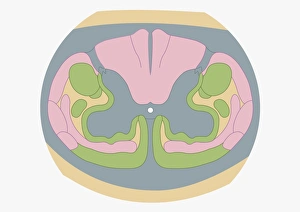
Normal axial view of C5 showing disk, nerve roots, posterior
Horizontal, Detail, Anatomy, Diagram, Illustration, Spine, Medical, Biology, Disk, Anterior, Posterior, Cut Out, White Background, Artwork, Color Image, Front View, Close Up, Healthcare And Medicine
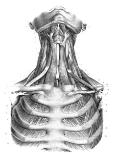
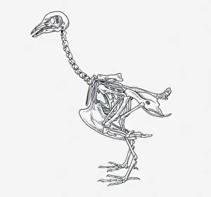
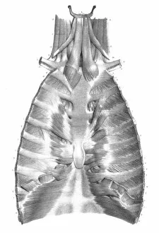
anatomy, animal themes, bird, coracoid, cranium, cut out, femur, humerous, mandible, no people, ostrich, pelvis girdle, pen and ink, phalanges, physiology, profile, pygostyle, side view, skeleton