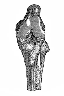
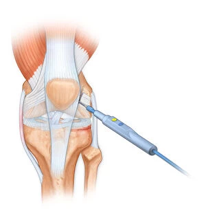
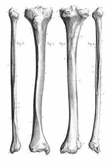
Bovie used to cut through retincaculum, and clean up femur of Displaced patellar knee
Detail, Anatomy, Diagram, Muscle, Illustration, Bone, Cutting, Equipment, Injury, Medical, Tool, Biology, Femur, Surgery, Knee, Torn, Fractured, Meniscus, Cut Out, White Background, Vertical
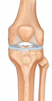
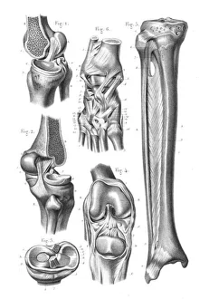
Illustration of the anterior knee, articular surface meniscus
anatomy, body part, bone, cartilage, close-up, color image, femur, fibula, front view, human body part, human bone, human joint, human knee, illustration, joint - body part, knee, patella, meniscus