mail_outline sales@mediastorehouse.com
1,278 Photographic Prints
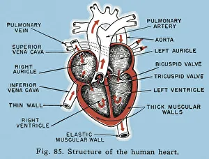
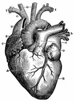
Human HeartAn anatomical diagram showing the arteries of the human heart, circa 1930. (Photo by Hulton Archive/Getty Images)
Labelled Structure of the Human Heart Diagram
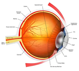
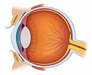
Human eye, illustrationEye anatomy. Cutaway illustration passing through a human eye, showing its internal anatomy and structure. The front of the eye is at right, and the structures here include the cornea
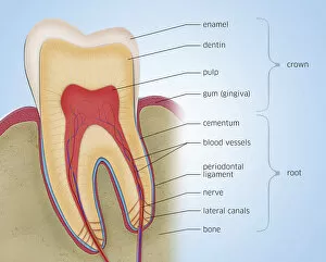
Tooth anatomy, illustrationCross-section of a molar tooth, illustration
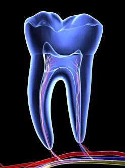
Molar toothTooth, transparent cross section of a molar tooth with arteries (red), veins (purple) and nerves (green)
Male blackcap (Sylvia atricapilla) perched on a pine tree branch. Photographed in Israel in October
Dental implants following tooth extraction, X-rayDental implants following tooth extraction, coloured X-ray. During this surgery, dental posts (white, centre) are placed in the jaw bone to allow a dental implant to be put in place to replace
Osteoarthritis of the cervical spine, X-rayOsteoarthritis of the cervical spine. Coloured lateral X-ray of the neck of a 76-year-old woman with severe osteoarthritis of the cervical spine
Surgical fixation for rheumatoid arthritis of feet, X-raySurgical fixation for rheumatoid arthritis of the feet. Coloured lateral X-ray of screws and pins used in surgical fixation of the foot joints of a 52-year-old woman with rheumatoid arthritis
Hip fracture, X-rayHip fracture. Frontal X-ray of the right hip of a 53-year-old man, showing a comminuted (splintered) fracture of the neck of the femur (centre left)
Fractured ankle bone, CT scanFractured ankle bone. Coloured computed tomography (CT) scan of the bones of the foot and ankle of a 23-year-old woman with a comminuted (splintered) fracture of the talus bone (centre) of the ankle
Arthritis of the neck, X-rayArthritis of the neck. Coloured X-ray of the arthritic cervical spine of a 70 year old man
Arthritis of the hip, X-rayArthritis of the hip. Coloured X-ray of an arthritic hip
Normal knee, X-rayNormal knee. Coloured X-ray of the knee of a 44 year old woman
Degenerative foot deformation, X-rayDegenerative foot deformation. Coloured X-ray of a section through the foot of a 66-year-old male patient with a severe degenerative change in the metatarsophalangeal (MTP)
Aspiration, chest X-rayAspiration. Coloured chest X-ray showing aspiration (dark areas) in the lungs of a 76-year-old female patient with an extensive brain haemorrhage
Lipoma of the shoulder, X-rayLipoma of the shoulder. Coloured X-ray of the shoulder of a 58-year-old male patient showing a large lipoma. Lipomas are benign (non-cancerous) tumours arising from adipose (fat) tissue
Tibial spur, X-rayTibial spur. Coloured X-ray of the foot of a 22-year-old male patient with a spur (osteophyte, highlighted) affecting the tibia (shin bone)
Pituitary tumour, CT scanPituitary tumour. Coloured computed tomography (CT) scan of a section through the brain of an 84-year-old male patient with a tumour (round, centre) affecting the pituitary gland
Pinned fractured hip, X-rayPinned fractured hip. Coloured X-ray of a section through the hip of a 91-year-old female patient with a fracture affecting the head of the femur (thigh bone, vertical)
Arthritic knees, X-rayArthritic knees. Coloured X-ray of the knees of a 66 year old male with osteoarthritis
Arthritis of the knee, X-rayArthritis of the knee. Coloured X-ray the arthritic knee of a 37 year old man. The knee had previously been damaged in a motorbike accident
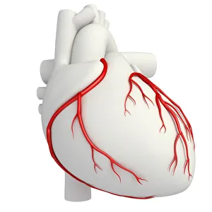
Coronary arteries, illustrationIllustration of coronary arteries of the heart
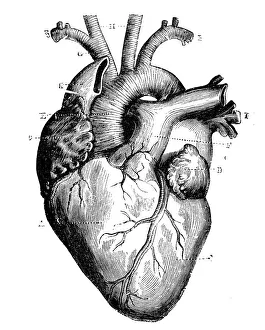
Antique medical scientific illustration high-resolution: heart
XXXL Very Detailed Human HeartEngraving From 1872 Featuring A Human Heart
Normal anatomy of the eye in cross sectionHorizontal, Vision, Detail, Anatomy, Diagram, Muscle, Illustration, Lens, Iris, Medical, Eyeball, Biology, Lateral, Cut Out, White Background, Eye, Artwork, Cross Section, Color Image, Close Up
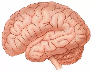
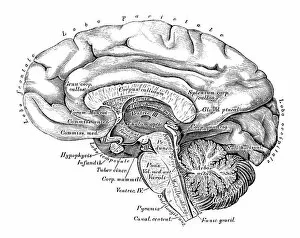
Lateral view of a normal brainHorizontal, Brain, Detail, Anatomy, Diagram, Illustration, Pink, Medical, Biology, Cut Out, White Background, Artwork, Color Image, Close Up, Healthcare And Medicine, The Human Body, No People
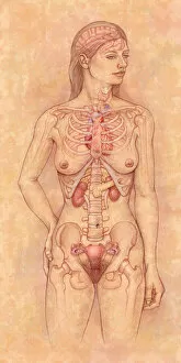
Front view of the female anatomy hilighting the endocrine systemBrain, Anatomy, Diagram, Heart, Illustration, Bone, Shoulder, Medical, Uterus, Biology, Pelvis, Femur, See Through, Vertical, Artwork, Female Likeness, Color Image, Front View
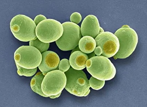
Yeast cells, SEMYeast cells. Coloured scanning electron micrograph (SEM) of cells of brewer s, or baker s, yeast (Saccharomyces cerevisiae). This fungus consists of single vegetative cells
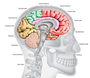
Human brain, illustrationIllustration of a cross-section of the brain showing the various lobes. The lobes are shown in different colours - red (frontal), green (parietal), yellow (occipital), orange (temporal)
Art, Asteroid, Astronomy, Black Hole, Colliding, Color Image, Concepts, CosmologyArt, Asteroid, Astronomy, Black Hole, Colliding, Color Image, Concepts, Science Photo Library, 85757958
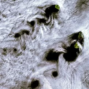
Bizarre, Climate, Cloud, Color Image, Destruction, Dramatic, Energy, EnvironmentBizarre, Climate, Cloud, Color Image, Destruction, Dramatic, Energy, En, Science Photo Library, 85758166
Astronomy, Brown, Color Image, Cosmology, Crater, Discovery, Exploration, GeologyAstronomy, Brown, Color Image, Cosmology, Crater, Discovery, Explorati, Science Photo Library, 85758294
Atmospheric, Chain, Climate, Cloud, Color Image, Directly Above, Energy, EnvironmentAtmospheric, Chain, Climate, Cloud, Color Image, Directly Above, Energ, Science Photo Library, 85758187
Human brain, illustrationIllustration of human brain
August, Bizarre, China, Climate, Cloud, Color Image, Cook Islands, Cyclone, DestructionAugust, Bizarre, China, Climate, Cloud, Color Image, Cook Islands, Cycl, Science Photo Library, 85757042
Astronomy, Black Background, Color Image, Concepts, Cosmology, Discovery, ExplorationAstronomy, Black Background, Color Image, Concepts, Cosmology, Discov, Science Photo Library, 85757371
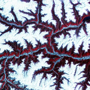
Color Image, Earth, Environment, February, Forest, Frozen, Geology, Himalayas, LandColor Image, Earth, Environment, February, Forest, Frozen, Geology, Him, Science Photo Library, 85757837
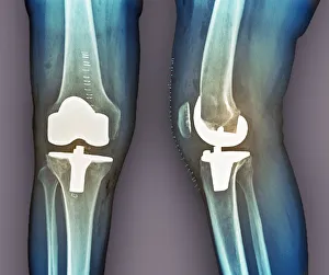
Total knee replacement, X-raysTotal knee replacement. Coloured frontal (left) and profile (right) X-rays of the right knee of a 69 year old patient after total knee replacement surgery
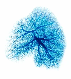
Lung, X-rayLung. Coloured X-ray showing the blood vessels in a lung
Human anatomy scientific illustrations: Brain side viewHuman anatomy scientific illustrations with latin/italian labels: Brain side view
Madagascar hissing cockroach (Gromphadorhina portentosa)
Mexican redknee tarantula (Brachypelma smithi)
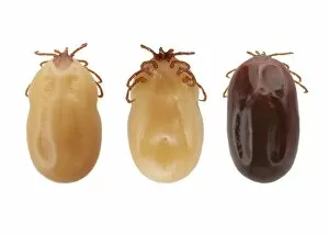
Ticks (superfamily Ixodoidea)
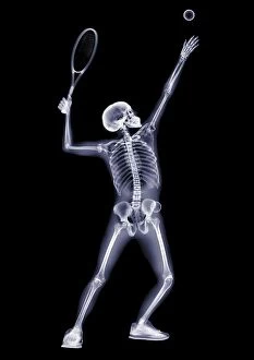
Person serving tennis ball, X-ray
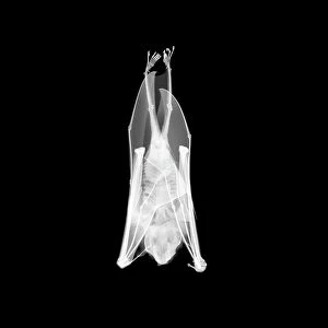
Flying fox, X-rayFlying fox, or fruit bat (Pteropus sp.), X-ray
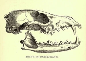
Skull of Canis magellanicus, 19th century illustration. From Dogs, Jackals, Wolves and Foxes. A Monograph of the Canidae, by George Mivart (F.R.S)
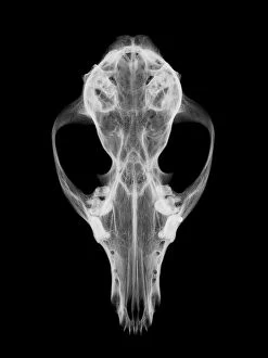
Fox skull, X-ray