mail_outline sales@mediastorehouse.com
128 items
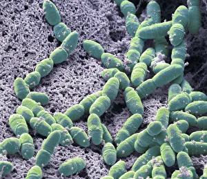
Streptococcus mutans, SEMStreptococcus mutans. Coloured scanning electron micrograph (SEM). S. mutans is a coccoid shaped, Gram-positive, anaerobic bacteria that is part of the normal bacteria flora of the mouth
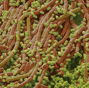
Plaque-forming bacteria, SEMPlaque-forming bacteria, coloured scanning electron micrograph (SEM). Plaque consists of a film of bacteria embedded in a glycoprotein matrix
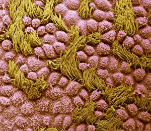
Fallopian tube, SEMFallopian tube. Coloured scanning electron micrograph (SEM) of the surface of a human fallopian tube. Fallopian tubes are ducts that lead from the ovaries to the uterus
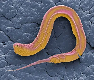
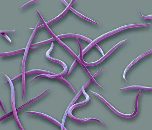
C elegans, SEMCaenorhabditis elegans worm, coloured scanning electron micrograph (SEM). C. elegans is a soil-dwelling hermaphrodite nematode worm and one of the most studied animals in biological
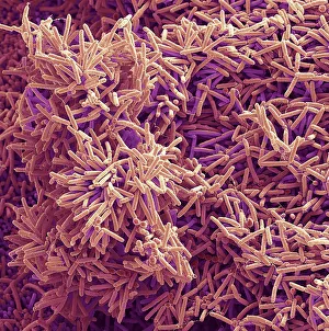
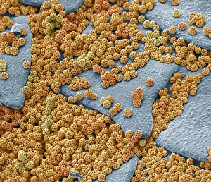
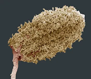
Soil bacteria, SEMSoil bacteria. Coloured scanning electron micrograph (SEM). Bacteria in the soil are directly tied to nutrient recycling especially carbon, nitrogen, phosphorus and sulfur
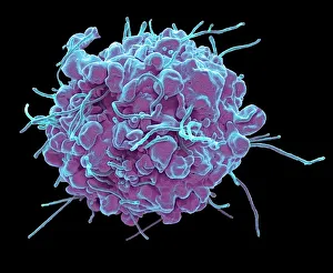
Resting T lymphocytes. Coloured scanning electron micrograph (SEM) of resting T lymphocytes from a human blood sample. T lymphocytes, or T cells
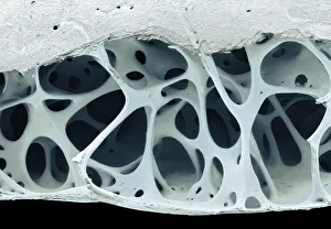
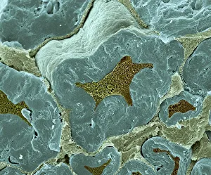
Bird bone tissue, SEMBird bone tissue. Coloured scanning electron micrograph (SEM) of cancellous (spongy) bone from a starlings (Sturnus vulgaris) skull
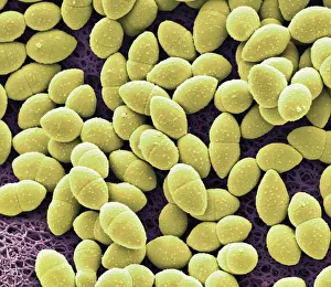
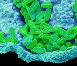
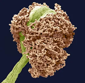
Kefir bacteria, SEMKefir bacteria. Scanning electron micrograph (SEM) of Lactococcus bacteria from kefir, a fermented milk beverage containing beneficial yeast as well as probiotic bacteria
Nematodes, SEMPhasmarhabditis hermaphrodita. Coloured scanning electron micrograph (SEM) of Phasmarhabditis hermaphrodita. Phasmarhabditis hermaphrodita is a microscopic nematode in the family Rhabditidae
Tendon, SEMTendon, coloured scanning electron micrograph (SEM), showing bundles of collagen fibres. The parallel alignment of the fibres make tendons inelastic but flexible. Tendons attach muscle to bone
Brochosomes, SEMBrochosomes, coloured scanning electron micrograph (SEM). Brochosomes are intricately structured microscopic granules secreted by leafhoppers
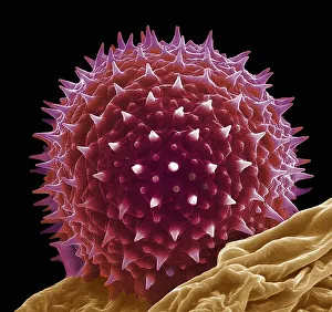
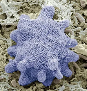
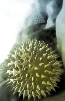
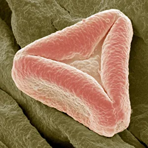
Verbena pollen, SEMVerbena pollen. Coloured scanning electron micrograph (SEM) of pollen grains from verbena bonariensis. Verbena bonariensis is a tall, slender-stemmed perennial
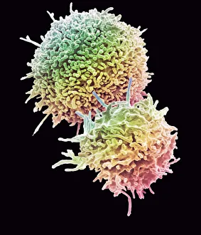
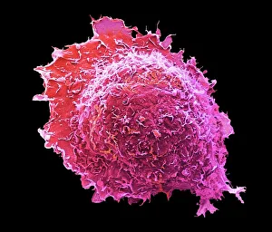
Apoptosis, SEMApoptosis. Coloured scanning electron micrograph (SEM) of a 293T cell in the early stages of programmed cell death, or apoptosis. Apoptosis occurs when a cell becomes old or damaged
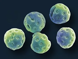
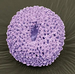
Diatoms, SEMDiatoms. Coloured scanning electron micrograph (SEM) of fresh water centric diatom frustules (skeleton), Diatoms are a type of algae (Chromophyta, Bacillariophyceae)
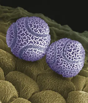
Convolvulus pollen grains, SEMConvolvulus pollen grains. Coloured scanning electron micrograph (SEM) of pollen grains from a convolvulus flower. Convolvulus is a genus of about 200 to 250 species of flowering plants in
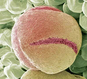
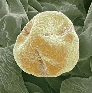
Hollyhock pollen grain, SEMHollyhock pollen grain. Coloured scanning electron micrograph (SEM) of a pollen grain from a hibiscus (Alcea setosa) flower. Pollen grains are the male sex cells of a flowering plant
Osteocyte, SEMOsteoblast bone cell. Coloured scanning electron micrograph (SEM) of an osteoblast bone cell. Osteocytes are osteoblasts (bone-producing cells) that have become trapped within bone cavities (lacunae)
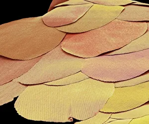
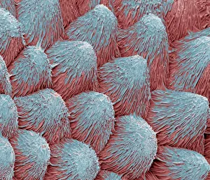
Silverfish scales, SEMSilverfish scales (Lepisma saccharina). Coloured scanning electron micrograph (SEM) of scales from a silverfish. The silverfish is a primitive insect that has remained unchanged for millions of
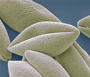
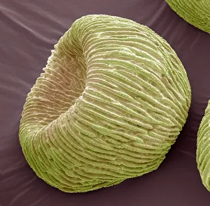
Water lily pollen grains, SEMWater lily pollen grains. Coloured scanning electron micrograph (SEM) of pollen grains from a water lily flower. Nymphaeaceae is a family of flowering plants, commonly called water lilies
Hibiscus pollen grains, SEMChinese hibiscus pollen. Coloured scanning electron micrograph (SEM) of pollen grains on the anther of a Chinese hibiscus (Hibiscus rosa-sinensis) flower
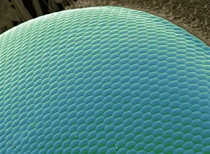
Wasp eye, SEMWasp eye. Coloured scanning electron micrograph (SEM) of numerous lenses, called ommatidia, that make up the compound eye of a wasp (Suborder Apocrita)
Phlox pollen, SEMPhlox pollen grain, magnified x2000 when printed at 10 centimetres wide
Bougainvillea pollen, SEMBougainvillea pollen grains, magnified x2000 when printed at 10 centimetres wide
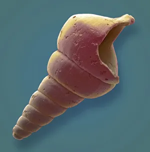
Gastropod microfossil, SEMGastropod. Coloured scanning electron micrograph (SEM) of a gastropod microfossil from maldives beach sand. Microfossils are roughly 0.05 to 2mm in size
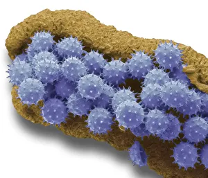
Korotnevella amoeba, SEMKorotnevella. Coloured scanning electron micrograph (SEM) of the scales covering a naked amoeba of the genus Korotnevella
Bellflower pollen, SEMBellflower pollen. Coloured scanning electron micrograph (SEM) of pollen grains from a bellflower (Campanula sp.). Pollen grains are the male gametes (sex cells) of a plant
Passion flower pollen. Coloured scanning electron micrograph (SEM) of pollen grains from a passion flower (Passiflora caerulea). Pollen grains are the male gametes (sex cells) of a plant
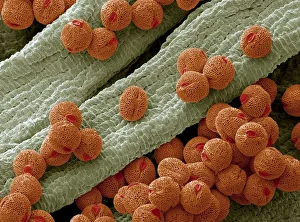
Asparagus pollen grains, SEMAsparagus (Asparagus officinalis) pollen grains, coloured scanning electron micrograph (SEM). Magnification: x2000 when printed at 10 centimetres wide
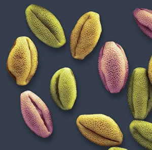
Electron Microscopic Image of Pollen GrainsA stylised scanning electron microscopic image of pollen grains. Pollen is a fine, coarse powdery substance comprising pollen grains which are male microgametophytes of seed plants
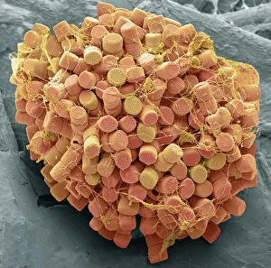
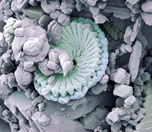
Fossil debris in chalk, SEMFossil debris in chalk. Coloured scanning electron micrograph (SEM) of a fossilized fragment of a coccolithotrope skeleton. Magnification: x8000 when printed at 10 centimetres wide
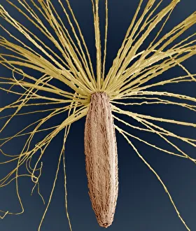
Smooth hawksbeard seed, SEMSmooth hawksbeard (Crepis capillaris) seed, magnified x35 when printed at 10 centimetres tall
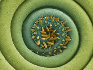
Bay tree anther, SEMStamen from the flower of a Bay Laurel tree (Laurus nobilis), magnified x70 when printed at 10 centimetres wide
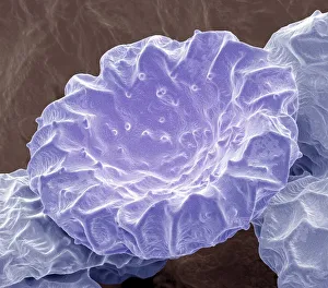
Wood anemone anther, SEMMale anther of a Wood Anemone (Anemone nemerosa) covered in pollen, magnified x65 when printed at 10 centimetres wide
Bottlebrush pollen, SEMBottlebrush (Callistemon) pollen grain, magnified x3000 when printed at 10 centimetres wide
Strawberry pollen, SEMStrawberry (Fragaria vesca) pollen grain, magnified x2000 when printed at 10 centimetres wide
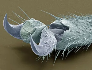
Stick insect foot, SEMClaw of a Stick Insect (Phasmatodea), magnified x80 when printed at 10 centimetres wide
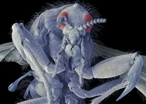
March fly, SEMMarch fly. Scanning electron micrograph (SEM) of a female march fly (family Bibionidae). A member of a family of stout insects in the fly order, Diptera
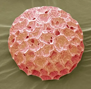
Orchid petal, SEMOrchid (Phalaenopsis) petal, magnified x450 when printed at 10 centimetres wide
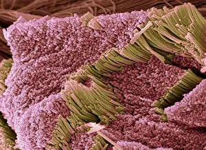
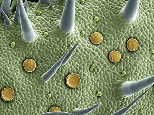
Marjoram leaf surface, scanning electron microscope (SEM)Backgrounds, Biology, Botany, Color Image, Full Frame, Glandular, High, SEM, 85758300
Geranium pollen, SEMGeranium pollen grain, magnified x650 when printed at 10 centimetres wide
Angels trumpets pollen, SEMAngels Trumpets (Brugmansias) pollen grain, magnified x1600 when printed at 10 centimetres wide
Kiwi fruit pollen grain, SEMkiwi fruit (Actinidia deliciosa) pollen grain, coloured scanning electron micrograph (SEM). Magnification: x3000 when printed at 10 centimetres wide
Scanning electron microscope (SEM) of tendonAnatomy, Biology, Close-Up, Color Image, Connective Tissue, Fiber, Ful, SEM, 85757662
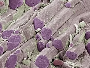
Skeletal muscle fibers, colored scanning electron micrograph (SEM)Anatomy, Backgrounds, Biology, Cartilage, Color Image, Connective Tis, SEM, 85757573
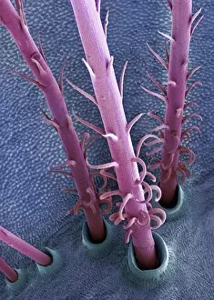
Caterpillar hairs, SEMCaterpillar hairs. Coloured scanning electron micrograph (SEM) of hairs from the vapourer moth (Orgyia antiqua) caterpillar. Magnification: x250 when printed at 10 centimetres tall
Moth proboscis. Coloured scanning electron micrograph (SEM) of the coiled proboscis of a moth (order Lepidoptera). The proboscis is an elongated part of the mouth
Nerve fibers, scanning electron micrographNerve fibres. Coloured scanning electron micrograph (SEM) of fractured myelinated nerve fibres. The myelin sheath is blue-green
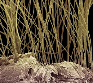
Dog hair, colored scanning electron micrograph (SEM)Dog hair, coloured scanning electron micrograph (SEM). The outside of the hair, the cuticle, is covered in overlapping scales of dead cells containing the protein keratin