mail_outline sales@mediastorehouse.com
Choose a picture from our Images Dated 9th July 2009 Collection for your Wall Art and Photo Gifts
12 items
Moth proboscis. Coloured scanning electron micrograph (SEM) of the coiled proboscis of a moth (order Lepidoptera). The proboscis is an elongated part of the mouth
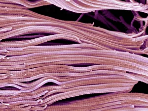
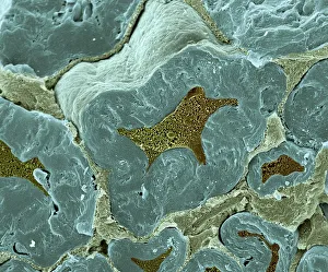
Collagen, Scanning electron micrograph (SEM)Collagen. Scanning electron micrograph (SEM) of collagen bundles from the delicate connective tissue endoneurium. Endoneurium wraps around and between individual nerve fibres (axons)
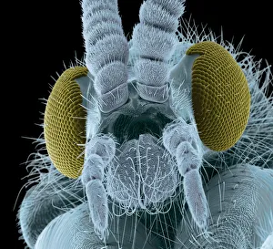
Fly head, colored scanning electron micrographFly head, coloured scanning electron micrograph (SEM). Close-up of the head of a fly, showing its short antennae (upper centre), which are seen between its compound eyes (brown)
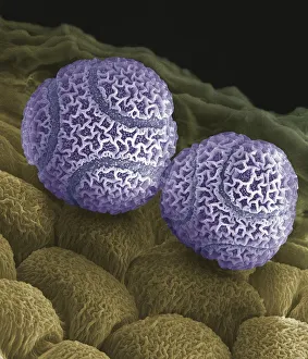
Passion flower pollen. Coloured scanning electron micrograph (SEM) of pollen grains from a passion flower (Passiflora caerulea). Pollen grains are the male gametes (sex cells) of a plant
Nerve fibers, scanning electron micrographNerve fibres. Coloured scanning electron micrograph (SEM) of fractured myelinated nerve fibres. The myelin sheath is blue-green
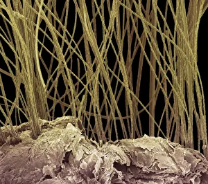
Dog hair, colored scanning electron micrograph (SEM)Dog hair, coloured scanning electron micrograph (SEM). The outside of the hair, the cuticle, is covered in overlapping scales of dead cells containing the protein keratin
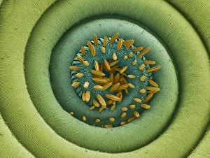
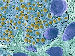
Pancreas tissue, colored scanning electron micrographPancreas tissue. Coloured scanning electron micrograph (SEM) of fractured pancreas tissue. Seen here are zymogen granules (yellow) and cell nuclei (purple)
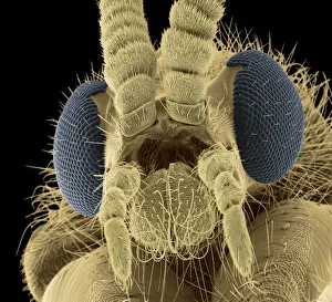
Fly head, colored scanning electron micrographFly head, coloured scanning electron micrograph (SEM). Close-up of the head of a fly, showing its short antennae (upper centre), which are seen between its compound eyes (blue)
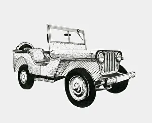
Black and white illustration of 1950s jeep
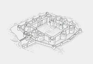
Black and white illustration of Beaumaris Castle
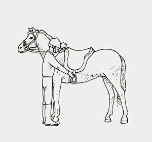
Black and white illustration of child preparing to get on horse
Old harry rocksVery low tide at Old Harry Rocks on world heritage UNESCO Jurassic coastline of Dorset, U.K