mail_outline sales@mediastorehouse.com
86 items
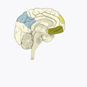
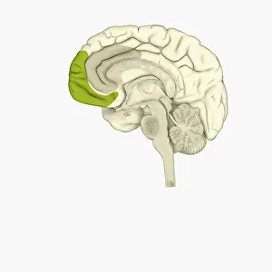
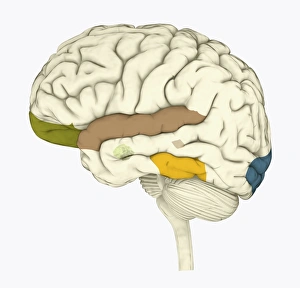
Digital illustration of posterior cingulate cortex (blue), medial frontal gyrus (yellow), and orbitofrontal prefrontal cortex (green) in human brain
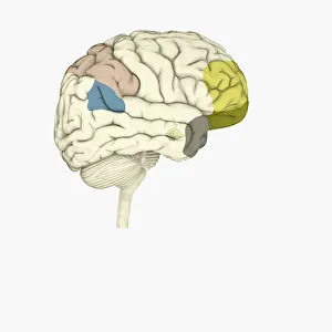
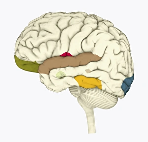
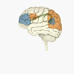
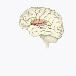
Digital illustration of parietal lobe (green), posterior superior temporal sulcas (blue), temperal pole (grey), dorsolateral prefrontal cortex, amygdala
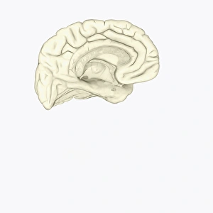
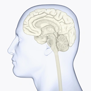
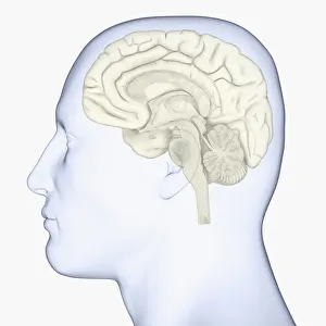
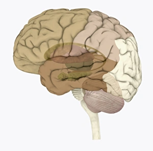
Digital cross section illustration of human brain
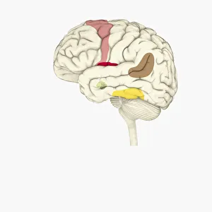
Digital illustration of emotional response areas in human brain
Digital illustration of anterior cingulate cortex (grey), and medial frontal cortex (green) in human brain
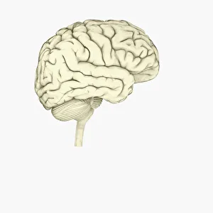
Digital illustration of human brain
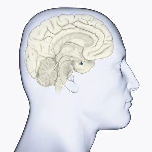
Digital illustration of head in profile showing pituitary gland in brain highlighted in blue
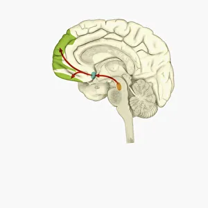
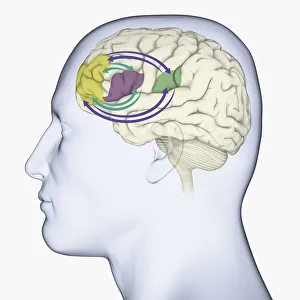
Digital illustration of reward pathway in human brain
Digital illustration of human brain associated with full awareness
Digital illustration of areas of information highlighted in human brain
Digital illustration of human brain with orbitofrontal cortex and amygdala highlighted in green
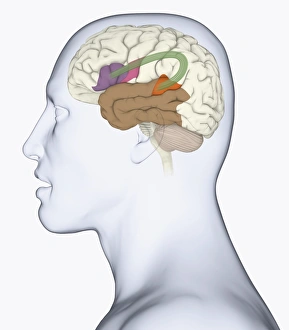
Digital illustration of head in profile showing areas of brain used for processing emotion
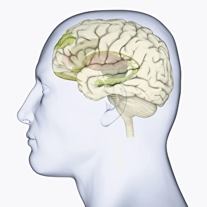
Digital illustration of head in profile showing brain
Digital illustration of head in profile showing direction of sensory signals from visual cortex to hippocampus in brain
Digital illustration of a tennis court with net in centre
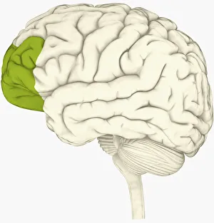
Digital illustration of prefrontal cortex of human brain highlighted in green
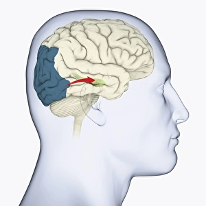
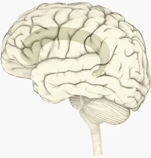
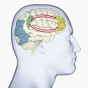
Digital illustration of frontal lobe and parietal lobe areas (orange) in left hemispheres (blue), and pathway of data from parietal lobe to frontal lobe (green) in human brain
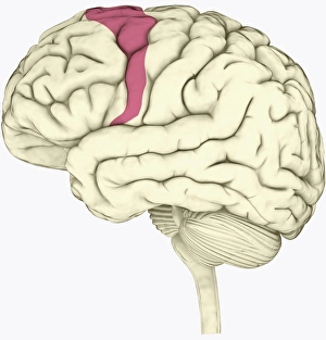
Digital illustration of premotor cortex involved in decision making highlighted in pink in human brain
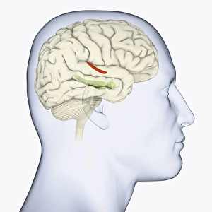
Digital illustration of head in profile showing hippocampus and amygdala (green), and auditory area (red) in brain
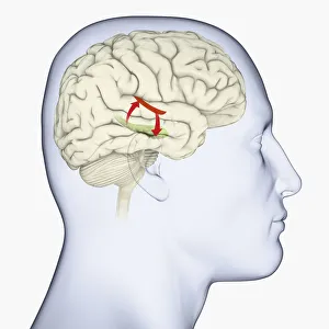
Digital illustration of head in profile showing auditory cortex (red), and hippocampus (green) and direction of signals in brain
Digital illustration of direction of dopamine flow, nucleus accumbens, basal ganglia and ventral tegmental area highlighted in human brain
Black and white digital illustration of cingulate cortex area of human brain
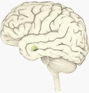
Digital illustration of amygdala in human brain
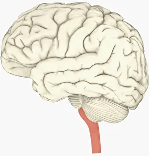
Digital illustration of human brain with spine highlighted in pink
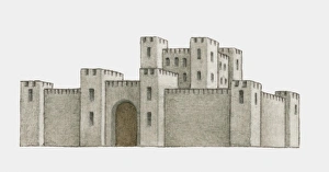
Illustration of 12th century northwestern European castle
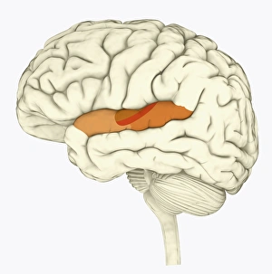
Digital illustration of human brain with primary auditory cortex highlighted in orange and red
Digital illustration of head in profile showing memory areas of brain
Digital illustration of head in profile showing amygdala, auditory cortex, wernickes area, and anterior temporal lobe in human brain
Digital illustration of head in profile showing bundle of nerve fibres connecti ng Brocas area and Wernickes area in human brain
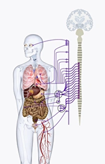
Digital illustration of autonomic nervous system responsible for automatic body functions
Digital illustration of areas associated with memory in human brain
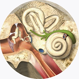
Digital illustration of middle and inner ear
Digital illustration of head in profile showing brain and right side of working memory
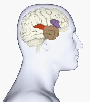
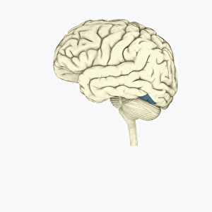
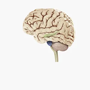
Digital illustration of hippocampus (green) and pons (blue) in left hemisphere of human brain
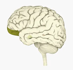
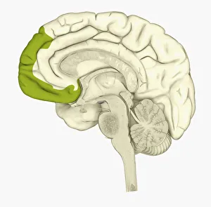
Digital illustration of human brain showing frontal cortex in green
Pumping WaterA domestic servant washing fish at the pump, circa 1857. From William Grundys English Views. (Photo by T. R. Williams/William Grundy/London Stereoscopic Company/Getty Images)