mail_outline sales@mediastorehouse.com
238 items
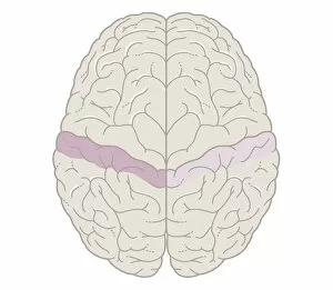
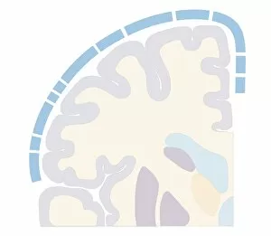
Cross section biomedical illustration of touch map of human brain
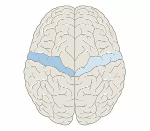
Cross section biomedical illustration of motor map of the brain
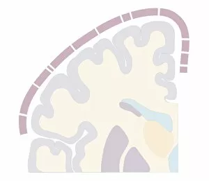
Cross section biomedical illustration of motor cortex topography of human brain
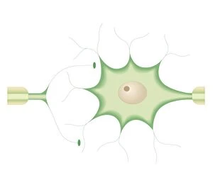
Cross section biomedical illustration of synapses between nerve cells
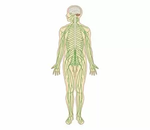
Cross section biomedical illustration of structure of the nervous system in adult male
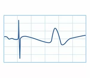
Biomedical illustration of nerve conduction trace
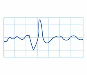
Biomedical illustration of electromyography (EMG) result
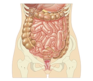
Cross section biomedical illustration of large and small intestine and pelvis in adult female
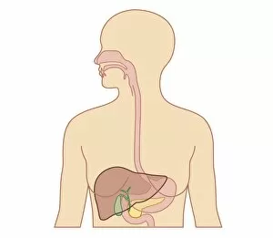
Cross section biomedical illustration of esophagus, liver, gallbladder, pancreas in adult female
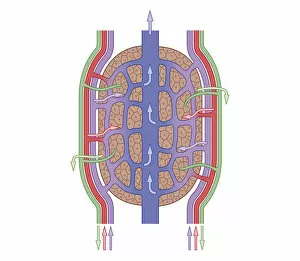
Cross section biomedical illustration of liver lobule
Cross section biomedical illustration of colonoscopy procedure in adult female
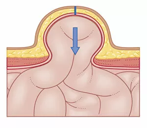
Cross section biomedical illustration of hernia repair and site of incision to uncover the hernia
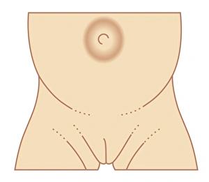
Biomedical illustration of umbilical hernia in female baby
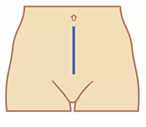
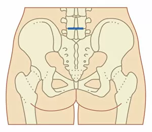
Cross section biomedical illustration of position of colectomy incision
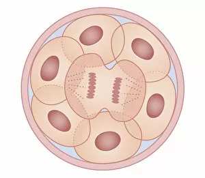
Cross section biomedical illustration of human cell division with zygote dividing to form new cells
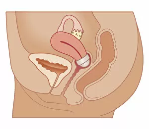
Cross section biomedical illustration of cervical cap in position
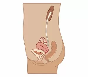
Cross section biomedical illustration of Intrauterine device (IUD) in position
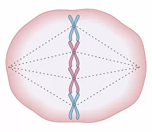
Cross section biomedical illustration of mitosis where the nucleus breaks down and threads form across cell with chromosomes lining up on these threads
Illustration of pie chart
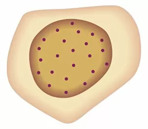
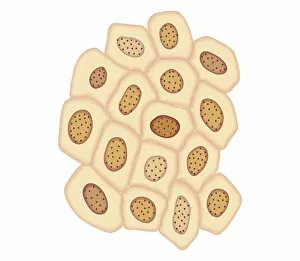
Cross section biomedical illustration of tumour-forming cancer cell
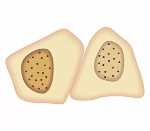
Cross section biomedical illustration of cancer cells forming tumour at first doubling stage
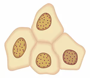
Cross section biomedical illustration of cancer cells forming tumour at second doubling stage
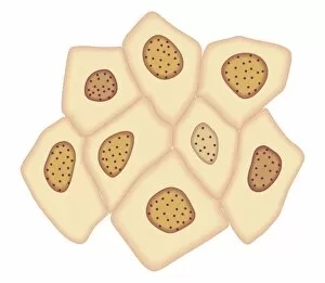
Cross section biomedical illustration of cancer cells forming tumour at third doubling stage
Cross section biomedical illustration of cancer cells forming tumour at fourth doubling stage
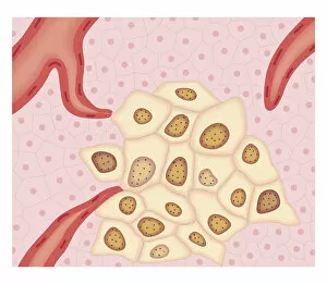
Cross section biomedical illustration of Angiogenesis - how tumour obtains nutrients
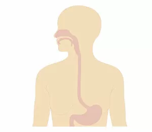
Cross section biomedical illustration of digestive tract in adult female
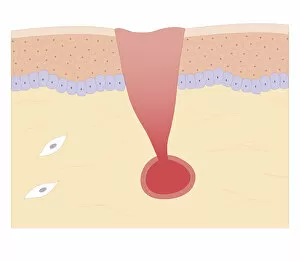
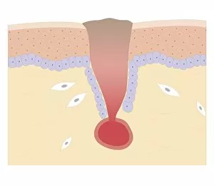
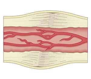
Cross section biomedical illustration of damaged skin and blood vessel
Cross section biomedical illustration of blood clotting over broken skin and fibroblasts multiplying and migrating to injury
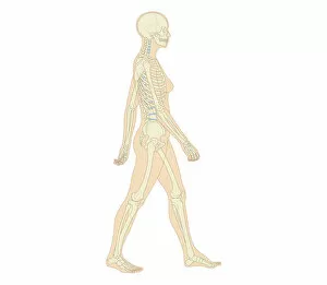
Cross section biomedical illustration of adult female human skeletal system and joints
Cross section biomedical illustration of lumber microdisectomy before surgery
Cross section biomedical illustration of lumber microdisectomy after surgery
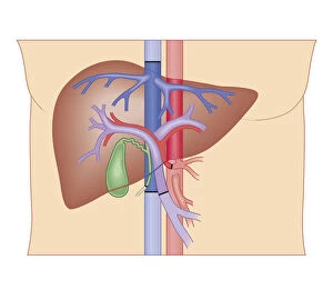
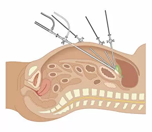
Cross section biomedical illustration of liver transplant procedure
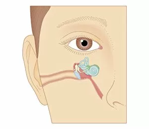
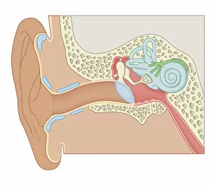
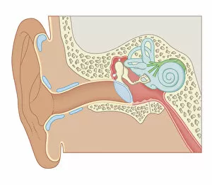
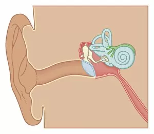
Cross section biomedical illustration of internal components of the ear
Cross section biomedical illustration of human ear
Cross section biomedical illustration of the anatomy of the ear
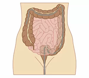
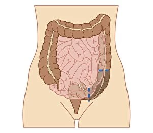
Cross section biomedical illustration of partial colectomy
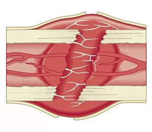
Cross section biomedical illustration of bone repairing itself with clot forming to seal broken blood vessels and fibrous tissue forming to replace the clot
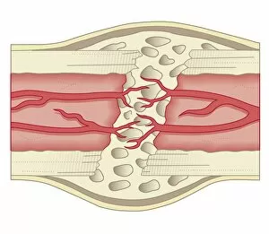
Cross section biomedical illustration of bone repairing itself with new, soft, spongy callus developing on framework provided by fibrous tissue, joining the broken ends
Cross section biomedical illustration of bone repairing itself with dense, compact bone gradually replacing the callus, and blood vessel regrowth
Cross section biomedical illustration of site of incision for microdiscectomy surgical procedure
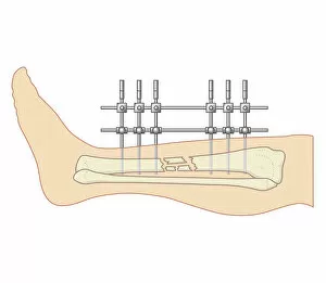
Cross section biomedical illustration of internal position external fixator used to repair broken leg
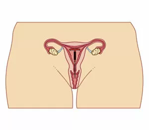
Cross section biomedical illustration of female urinary and reproductive system in adult female
Cross section biomedical illustration of endoscope and tubes inserted in abdomen of adult female during Laparoscopic Surgery
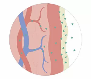
Cross section biomedical illustration of nasal / sublingual / buccal route with drugs absorbed through thin mucusCross section biomedical illustration of nasal/sublingual/buccal route with drugs absorbed through thin mucus membrane directly into bloodstream
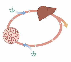
Cross section biomedical illustration of how oral and non-oral drugs circulate in the body, showing liver and inset
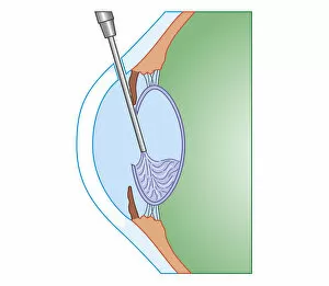
Cross section biomedical illustration of eye cataract surgery sucking softened tissue out using ultrasound probe inserted into lens via incision in cornea