
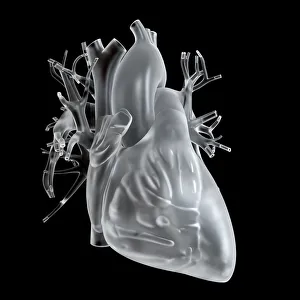
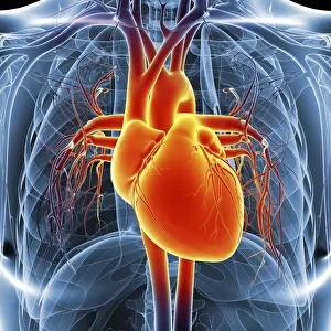
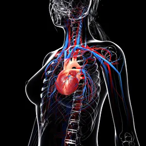
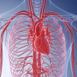
A stunning tribute to the intricate beauty of one of the most vital organs in the human body
Choose a picture from our A stunning tribute to the intricate beauty of one of the most vital organs in the human body Collection for your Wall Art and Photo Gifts
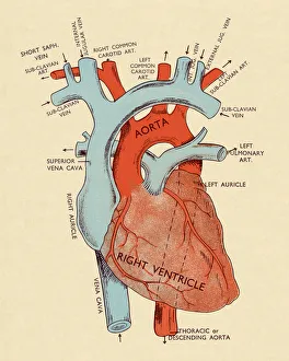
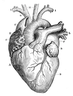
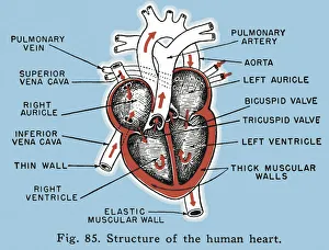
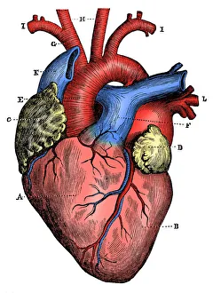
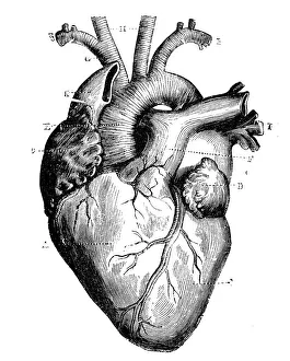
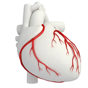
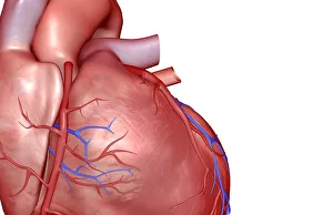
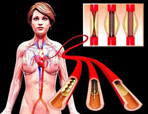
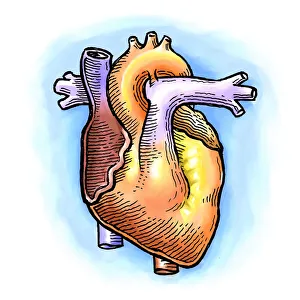
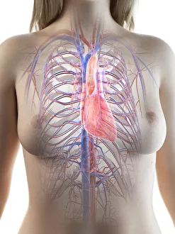
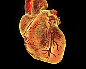
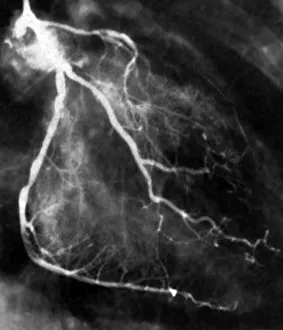
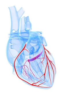
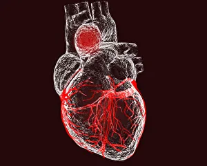
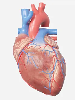
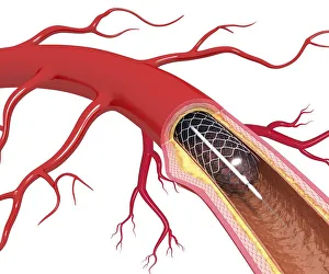
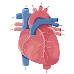
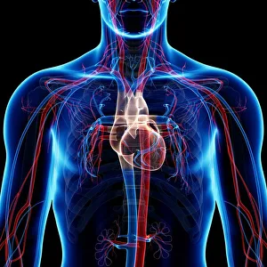
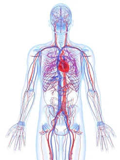
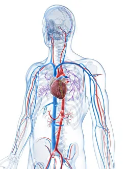
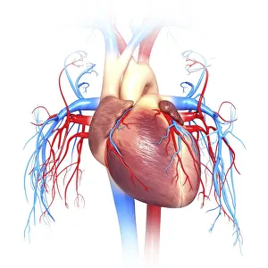
Our collection of artistic interpretations of the human heart, from anatomically correct depictions to abstract representations, is inspired by scientific wonders. Each image exemplifies the complexity and elegance of this magnificent instrument, capturing its beauty and significance in both literal and metaphorical ways.
106 items

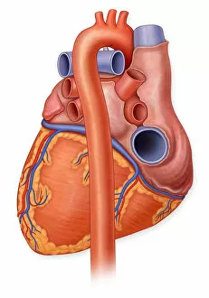
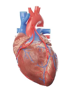
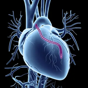
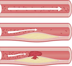
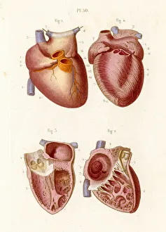

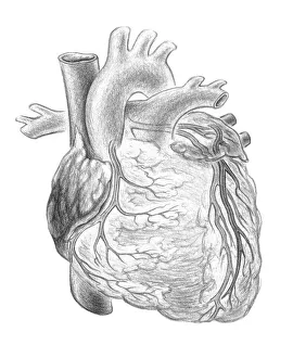
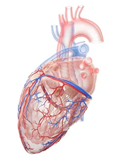
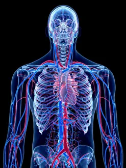
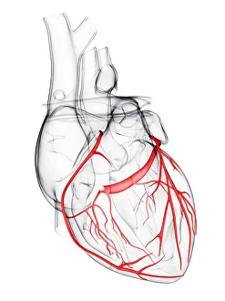
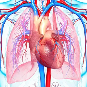


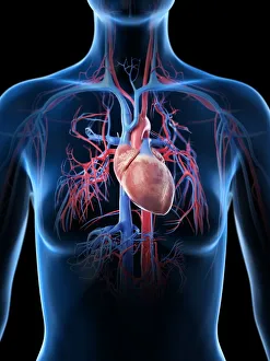
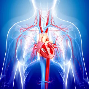
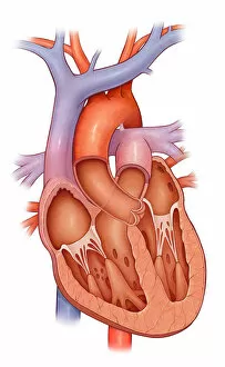

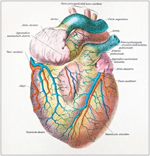
Our human heart collection is sure to captivate and inspire you, whether you are a medical professional, a science enthusiast, or simply appreciate the beauty of the human body.