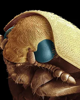
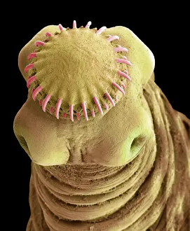
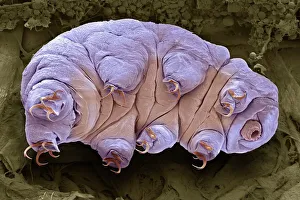
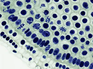
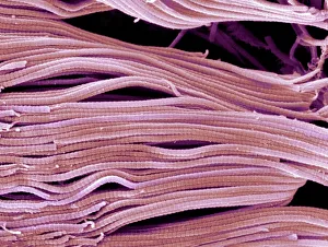
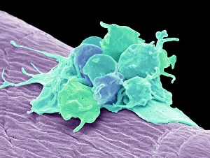
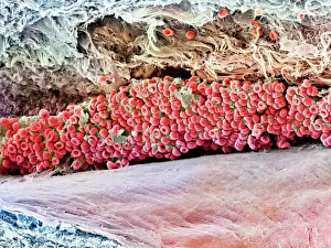
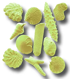
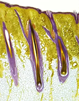
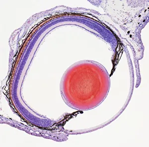
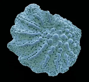
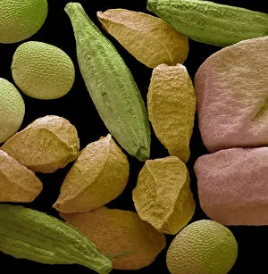
SEM (Scanning Electron Microscope) Inspired Art Collection
Welcome to the world of tiny creatures and objects around us beautifully photographed under an electron microscope
Choose a picture from our SEM (Scanning Electron Microscope) Inspired Art Collection for your Wall Art and Photo Gifts
128 items