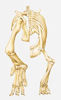
Illustration of the lower skeleton of an Euparkeria, a theocodont archosaur, Early Triassic period
animal foot, animal leg, animal skeleton, archosaur, dinosaur, euparkeria, extinct, low section, no people, palaeontology, prehistoric era, studio shot, theocodont, vertical, watercolour painting
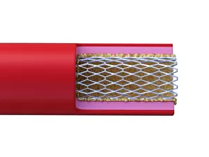
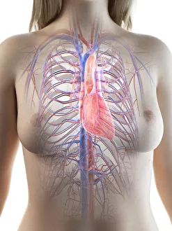
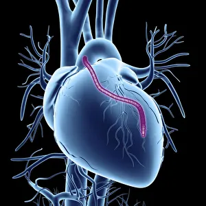
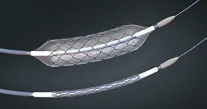
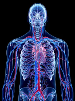
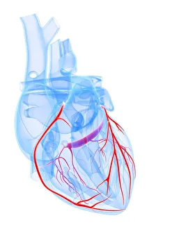
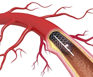
Angioplasty, computer artwork
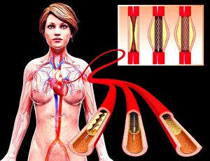
one person:CB2, atherosclerosis:CB2, repairing:CB2, women:CB2, half-length:CB2, atheroma:CB2, heart disease:CB2, surgery:CB2, black background:CB2, heart:CB2, front view:CB2, aorta:CB2
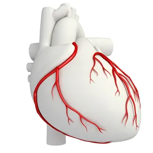
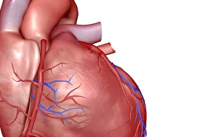
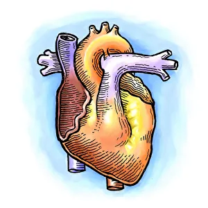
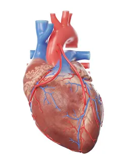
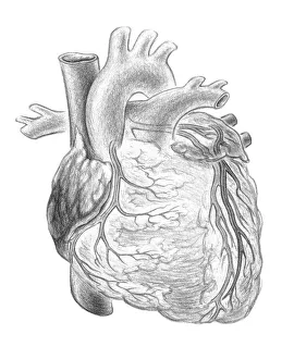
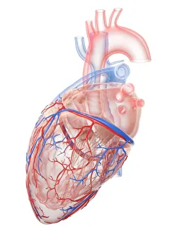
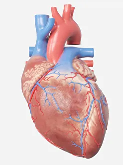
Posterior view of a normal heart and its arteries
Detail, Anatomy, Diagram, Muscle, Heart, Illustration, Medical, Biology, Cardiac, Rear View, Cardiovascular, Cut Out, White Background, Vertical, Artwork, Color Image, Close Up
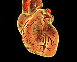
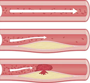
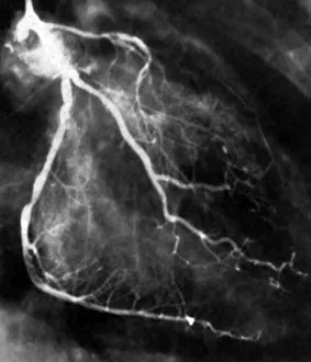
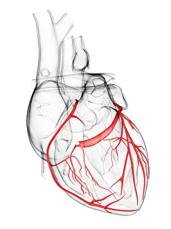
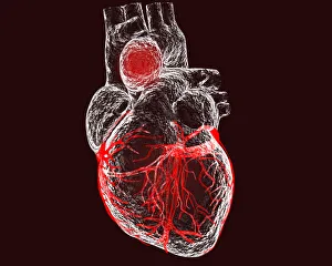
Narrowed coronary arteries (B&W)
Anatomy, Black And White, Block, Cardiovascular System, Chest, Coronary Artery, Healthcare And Medicine, Human Artery, Human Heart, Medicine, Monochrome, Narrow, Patient, Photography, Radiologist