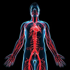
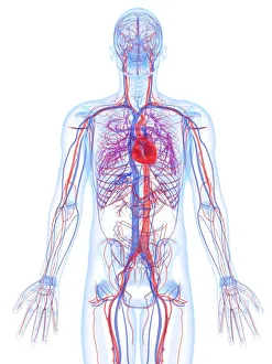
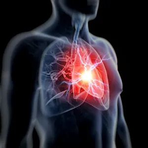
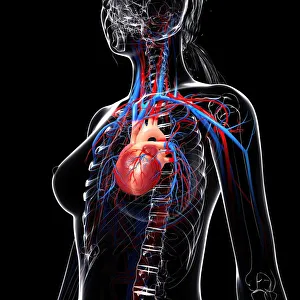
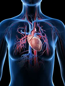
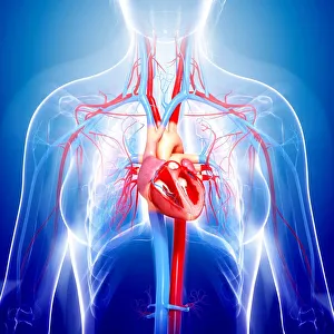
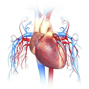
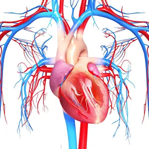
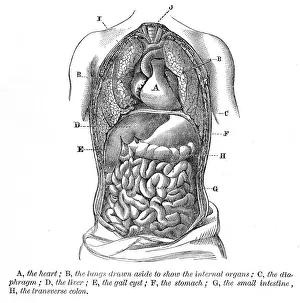
Female cardiovascular system, computer artwork
one person:CB2, half-length:CB2, artery:CB2, veins:CB2, anatomy:CB2, black background:CB2, view from below:CB2, three-quarter view:CB2, female:CB2, translucent:CB2, heart:CB2, human body:CB2
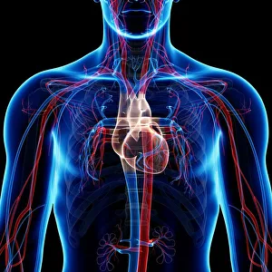
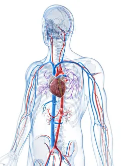
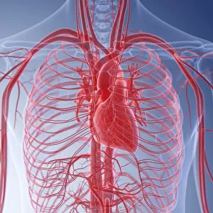
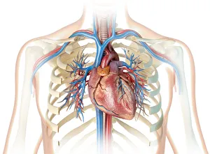
Female cardiovascular system, computer artwork
torso:CB2, one person:CB2, women:CB2, half-length:CB2, veins:CB2, anatomy:CB2, blue background:CB2, translucent:CB2, heart:CB2, front view:CB2, aorta:CB2, carotid artery:CB2, human body:CB2
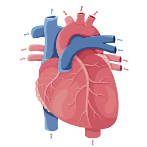
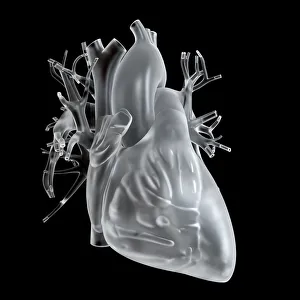
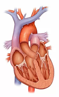
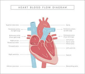
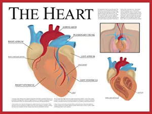
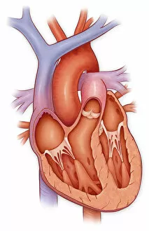
Cross section of a normal heart
Detail, Anatomy, Diagram, Muscle, Heart, Illustration, Biology, Chamber, Cardiac, Atrium, Cardiovascular, Cut Out, White Background, Vertical, Artwork, Cross Section, Color Image, Close Up
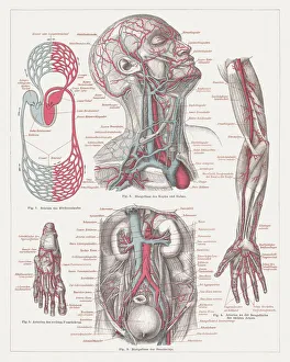
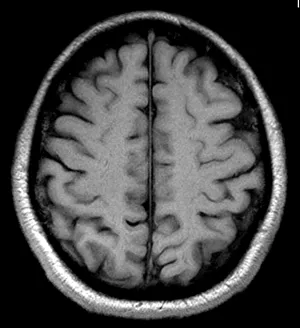
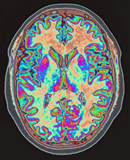
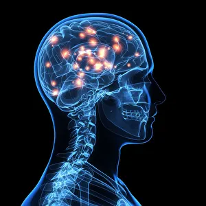
MRI scan of brain
body part, brain, bright, colour, conditions, medical, medical scan, medical treatment, mri scan, multi coloured, one person, someone, technology, vertical, photography, brain artwork, 56961661
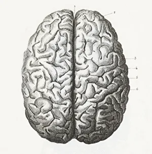
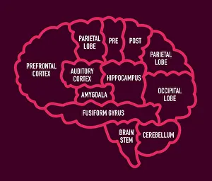
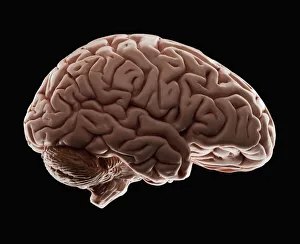
Model of human brain, studio shot
intelligence, anatomical model, anatomy, black background, brain, complexity, healthcare and medicine, horizontal, no people, pattern, single object, studio shot, wisdom, brain artwork, 114850733
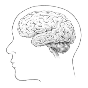
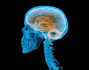
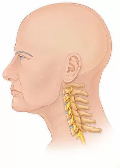
Normal lateral view of a mans head and neck with a skull
Anatomy, Diagram, Illustration, Spine, Medical, Neck, Biology, Disks, Disk, See Through, Cut Out, White Background, Profile, Vertical, Artwork, Male Likeness, Color Image, Healthcare And Medicine
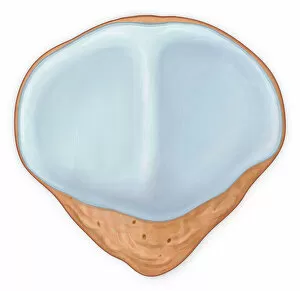
Posterior view patellar surface showing normal cartilage
Horizontal, Detail, Anatomy, Diagram, Illustration, Bone, Medical, Biology, Rear View, Cut Out, White Background, Artwork, Color Image, Close Up, Healthcare And Medicine, The Human Body, No People
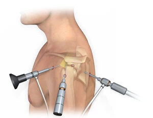
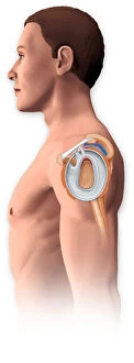
Lateral view of arthroscopic surgical repair on the shoulder joint
Horizontal, Detail, Anatomy, Diagram, Repair, Illustration, Bone, Shoulder, Equipment, Medical, Tool, Biology, Surgery, Joint, See Through, Cut Out, White Background, Artwork, Male Likeness
Open shoulder joint showing inflamed bursa from a bone spur and torn labrum
Detail, Anatomy, Diagram, Illustration, Bone, Shoulder, Injury, Medical, Biology, Joint, Torn, Injured, Cut Out, White Background, Vertical, Artwork, Color Image, Close Up, Healthcare And Medicine
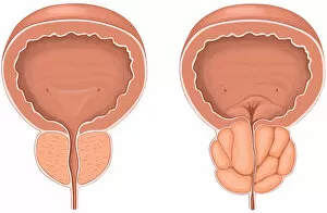
Anterior view showing normal versus enlarged prostate gland
Horizontal, Detail, Anatomy, Diagram, Illustration, Contrast, Medical, Biology, Abnormal, Anterior, Cut Out, White Background, Artwork, Cross Section, Color Image, Front View, Close Up
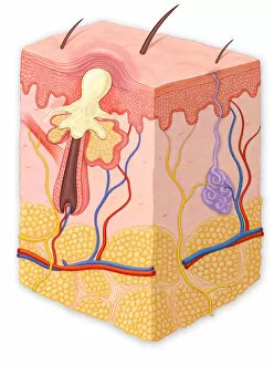
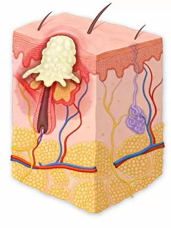
Skin cross section showing a papule
Detail, Anatomy, Diagram, Illustration, Medical, Vein, Skin, Biology, Fat, Epidermis, Cut Out, White Background, Hair, Vertical, Artwork, Cross Section, Color Image, Close Up
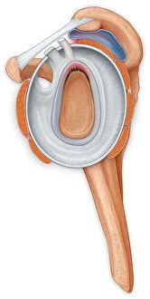
Lateral view of the shoulder joint showing a tear in the labrum and an inflamed bursa
Anatomy, Diagram, Illustration, Shoulder, Injury, Medical, Biology, Surgery, Joint, See Through, Cut Out, White Background, Vertical, Artwork, Male Likeness, Color Image, Healthcare And Medicine
Lateral view of the shoulder joint showing the repair to the torn labrum
Anatomy, Diagram, Repair, Illustration, Shoulder, Injury, Medical, Biology, Surgery, Joint, Torn, See Through, Cut Out, White Background, Vertical, Artwork, Male Likeness, Color Image
Skin cross section showing a pustule
Detail, Anatomy, Diagram, Illustration, Medical, Vein, Skin, Biology, Fat, Veins, Epidermis, Cut Out, White Background, Hair, Vertical, Artwork, Cross Section, Color Image, Close Up
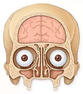
Normal coronal section of the skull and brain showing the coronal sinuses
Brain, Anatomy, Diagram, Illustration, Bone, Skull, Medical, Eyeball, Biology, See Through, Cut Out, White Background, Eye, Vertical, Artwork, Color Image, Front View, Human Skull
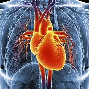
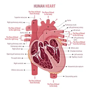
Cross section of a normal heart
Detail, Anatomy, Diagram, Muscle, Heart, Illustration, Medical, Biology, Atrium, Cardiovascular, Cut Out, White Background, Vertical, Artwork, Cross Section, Color Image, Close Up
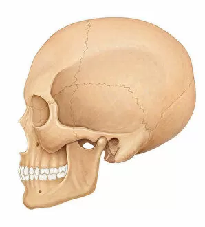
Normal lateral view of an adult skull
Anatomy, Diagram, Illustration, Bone, Teeth, Skull, Medical, Biology, Side View, Cut Out, White Background, Profile, Vertical, Artwork, Color Image, Human Skull, Healthcare And Medicine
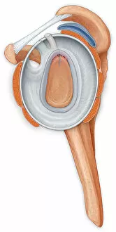
Lateral view of the shoulder joint showing the repair to the labrum
Detail, Anatomy, Diagram, Muscle, Illustration, Bone, Shoulder, Medical, Biology, Joint, Cut Out, White Background, Vertical, Artwork, Color Image, Close Up, Healthcare And Medicine, The Human Body