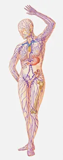
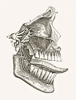
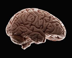
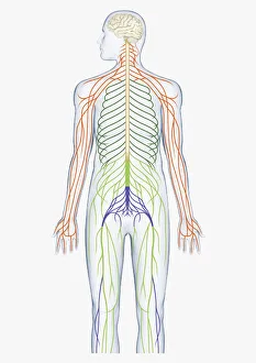
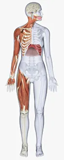
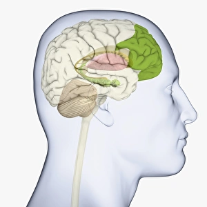
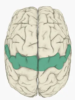
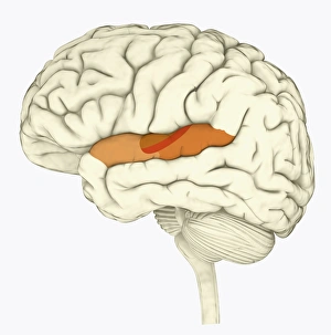
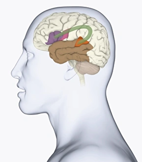
Anatomical Model Collection
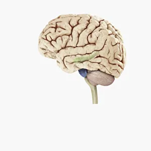
Model of human brain, studio shot
intelligence, anatomical model, anatomy, black background, brain, complexity, healthcare and medicine, horizontal, no people, pattern, single object, studio shot, wisdom, brain artwork, 114850733
All Professionally Made to Order for Quick Shipping