mail_outline sales@mediastorehouse.com
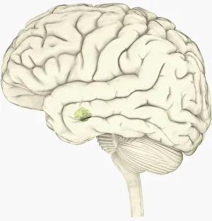
Digital illustration of amygdala in human brain
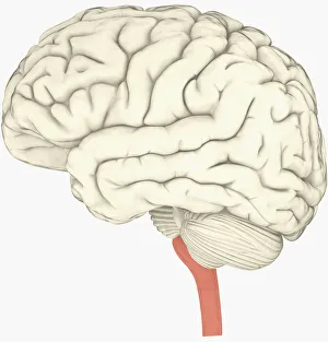
Digital illustration of human brain with spine highlighted in pink
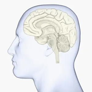
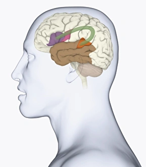
Digital illustration of head in profile showing brain
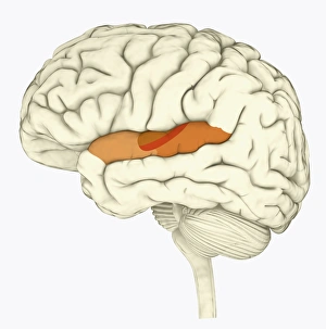
Digital illustration of human brain with primary auditory cortex highlighted in orange and red
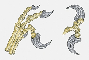
Illustration of Deinonchus foot showing large talons
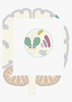
Digital illustration of basal ganglia
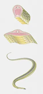
Sequence of illustrations showing Paradise Tree Snake (Chrysopelea paradisi) and anatomy cross sections
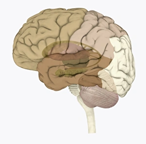
Digital illustration of head in profile showing memory areas of brain
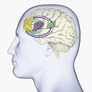
Digital illustration of head in profile showing bundle of nerve fibres connecti ng Brocas area and Wernickes area in human brain
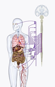
Digital illustration of autonomic nervous system responsible for automatic body functions
Digital illustration of areas associated with memory in human brain
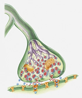
Cross section illustration of Synapse
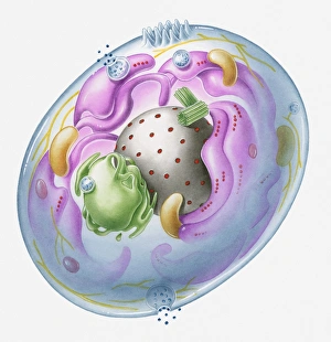
Cross section illustration of generalised human cell with major organelles
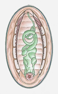
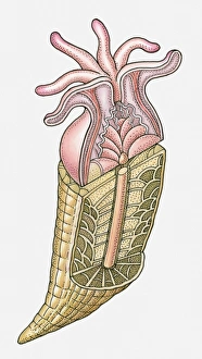
Anatomical illustration of a Chiton
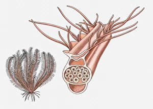
Illustration of a Feather star (Crinoidea) and its reproductive pinnule
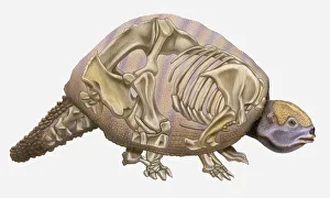
Anatomical illustration of a Pleistocene Edentate (Glyptodon reticulatus), an early mammal
Anatomical illustration of an Ammonite
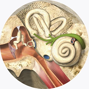
Digital illustration of middle and inner ear
Anatomical illustration of a coral
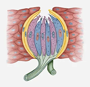
Cross section illustration of human taste bud
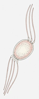
Illustration of an Eukaryote cell
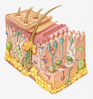
Cross section illustration of human skin showing touch receptor nerves
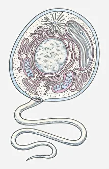
Illustration of a Prokaryote cell
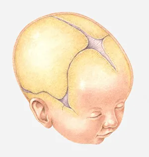
Illustration of Coronal Suture on head of newborn baby
Anatomical illustration of an Euparkeria, a thecodont archosaur, Triassic period
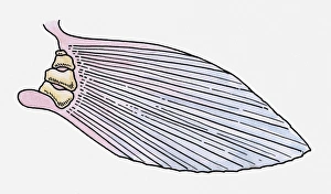
Illustration of the fin of a prehistoric Ray-finned fish (Actinopterygii)
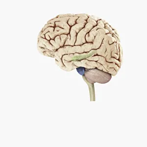
Digital illustration of hippocampus (green) and pons (blue) in left hemisphere of human brain
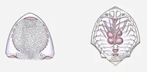
Anatomical illustration of a Cephalaspid prehistoric fish, underside of head and internal head structure
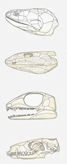
Illustration of the skulls of four vertebrates, fleshy-finned fish (Sarcopterygii), early tetrapod, early reptile, mammal
Sequence of illustrations showing complete bone fracture and healing process
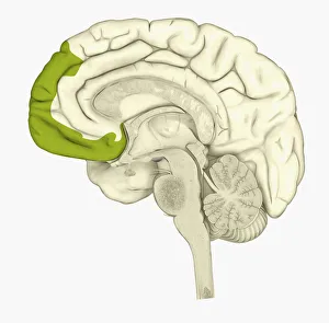
Digital illustration of human brain showing frontal cortex in green
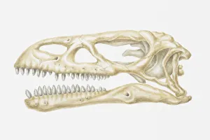
Illustration of the skull of a Scelidosaurus, a type of Thyreophoran dinosaur, Jurassic period
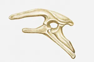
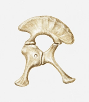
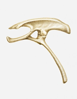
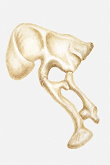
Illustration of the hip bone of a Stegosaurus, a type of Thyreophoran dinosaur, Jurassic period
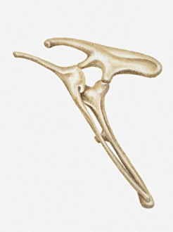
Illustration of the hip bones of a Hypsilophodon, a type of Ornithopod dinosaur, early Cretaceous period
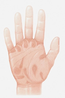
Illustration of accu-pressure points on palm of human hand
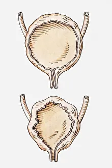
Illustration of full and empty human bladder
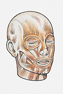
Illustration showing human head muscles
Illustration of the hip bone of a Diplodocus dinosaur, Jurassic period
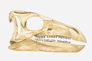
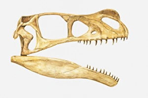
Illustration of the skull of an Ornitholestes, a theropod dinosaur from the Jurassic period
Illustration of the skull of a Stegosaurus, a type of Thyreophoran dinosaur, Jurassic period
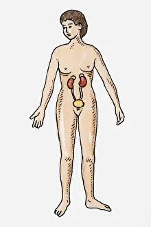
Illustration of female urinary system
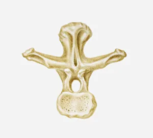
Illustration of the vertebra of a Brachiosaurus, late Jurassic-Cretaceous periods
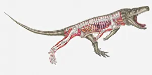
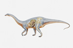
Illustration of the internal anatomy of a Riojasaurus, Triassic period
Illustration of the hip bone of a Homalocephale dinosaur, late Cretaceous period
Illustration of the hip bone of a Segnosaurus, a bipedal dinosaur, Cretaceous period
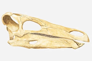
Illustration of the skull of a Massospondylus dinosaur, Jurassic period
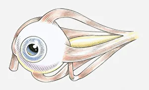
Illustration of muscles attached to human eye
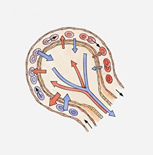
Illustration showing gas exchange in alveolus