mail_outline sales@mediastorehouse.com
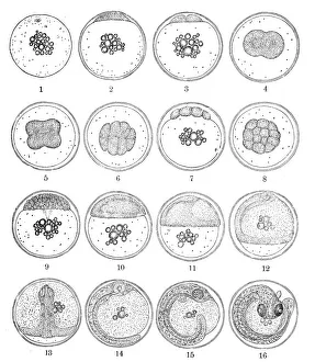
The three-spined stickleback (Gasterosteus aculeatus) egg developmentIllustration of a The three-spined stickleback (Gasterosteus aculeatus) egg development
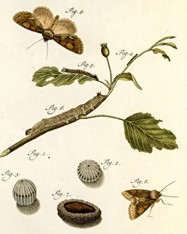
Metamorphosis, 19 century science illustrationMetamorphosis of insects. A photo of an original hand-colored engraving from Johann Christiaan Seppas Beschouwing der wonderen Gods, in de minst geachte schepzelena published in 1762
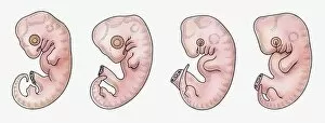
Illustration of reptile, bird, rabbit and human embryos in early stage of development
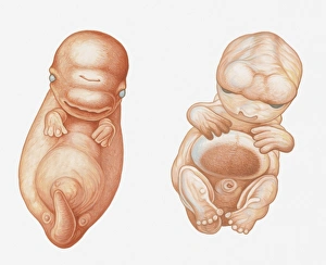
Illustration of Pacific White-sided Dolphin (Lagenorhynchus obliquidens) embryo at seven weeks, and human embryo (Homo sapiens) at seven weeks
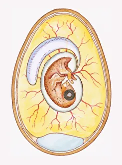
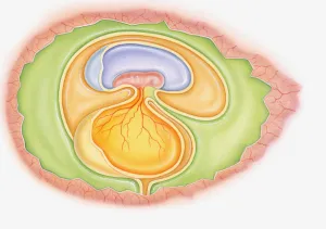
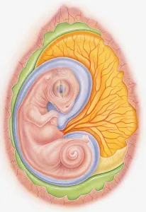
Illustration of yoke sac and amniotic membrane surrounding chicken embryo
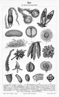
Eggs of fish and lower animals engraving 1895Meyers Konversations-Lexikon. Ein Nachschlagewerk des allgemeinen Wissens, 5th edition 17 volumes Bibliographisches Institut - Leipzig 1895-1897
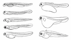
Various fish embryosIllustration of a Various fish embryos
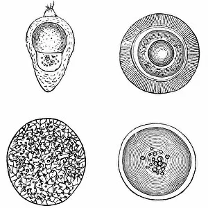
Various fish eggsIllustration of a various fish eggs
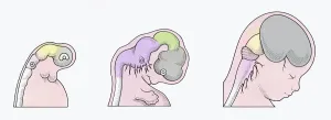
Illustration of three stages of Neurogenesis responsible for populating growth of brain in human embryo
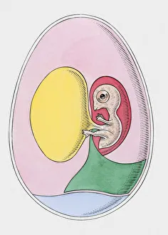
Illustration of dinosaur foetus in egg showing yoke sac and amniotic membrane
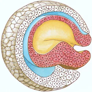
Illustration of cross section showing how early stage embryo of Flatworm (Phylum Platyhelminthes) differentiates into three layers of cell tissue, ectoderm, mesoderm and endoderm
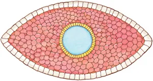
Illustration of cross-section of embryo forming the blastophore
Illustration of dinosaur embryo in egg showing yoke sac and amniotic membrane
Cross section illustration of bird embryo inside egg