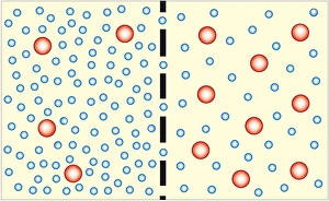
Biology Collection (page 18)

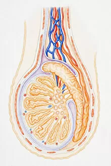


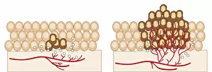
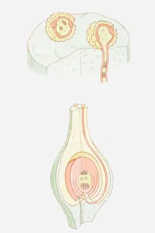
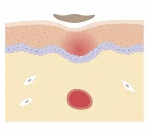
Digital illustration of angiogenesis process showing dormant tumour invading tissue, growing to mali
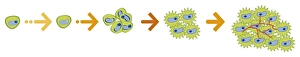
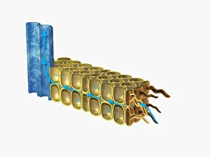
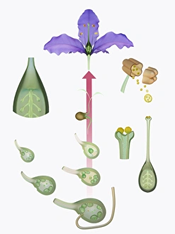
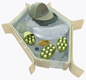
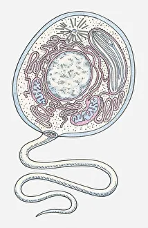
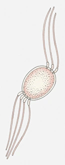
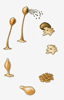
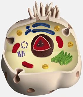
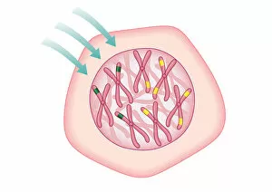
Illustration of the structure of a plant cell, including nucleus, nucleolus, ribosome, endoplasmatic
Digital illustration of angiogenesis process showing dormant tumour invading tissue, growing to mali
Illustration of the structure of a plant cell, including nucleus, nucleolus, ribosome, endoplasmatic