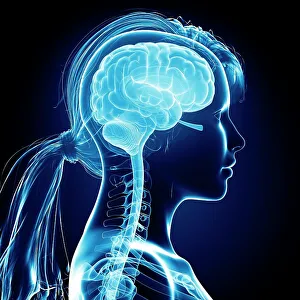
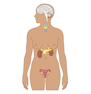
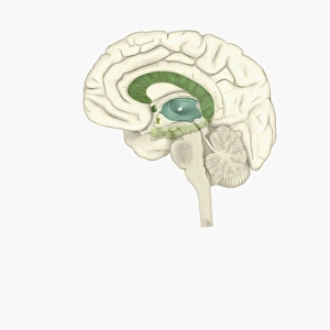
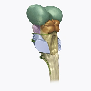
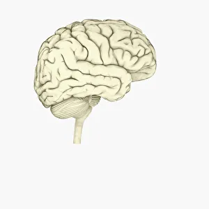
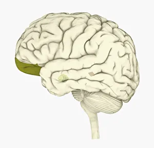
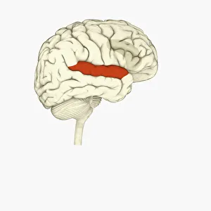
Female brain, computer artwork
cerebellum:CB2, cerebrum:CB2, spinal column:CB2, adult:CB2, head and shoulders:CB2, side view:CB2, anatomy:CB2, illustration:CB2, black background:CB2, female:CB2, translucent:CB2, nobody:CB2
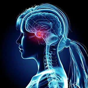
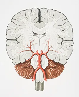
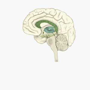
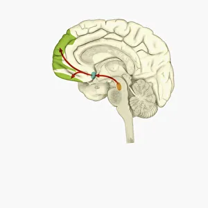
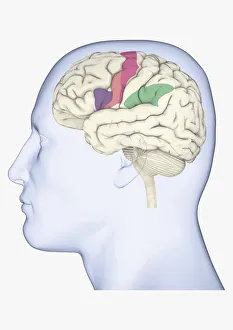
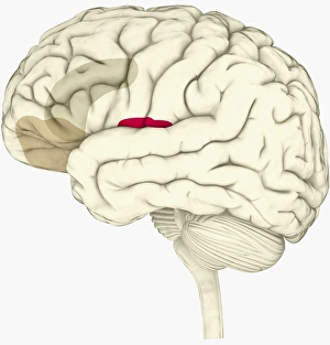
Female brain, computer artwork
cerebellum:CB2, cerebrum:CB2, spinal column:CB2, head and shoulders:CB2, side view:CB2, anatomy:CB2, illustration:CB2, black background:CB2, female:CB2, translucent:CB2, pituitary gland:CB2
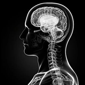
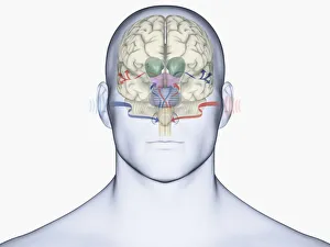
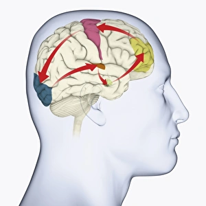
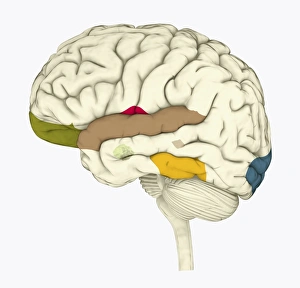
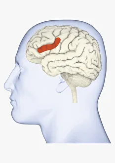
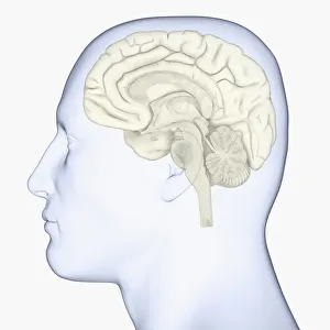
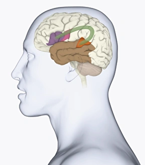
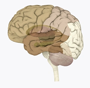
Male brain, computer artwork
cerebellum:CB2, cerebrum:CB2, adult:CB2, head and shoulders:CB2, side view:CB2, visual arts:CB2, anatomy:CB2, illustration:CB2, black background:CB2, male:CB2, translucent:CB2, nobody:CB2
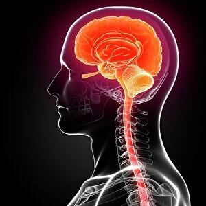
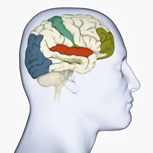
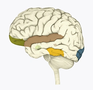
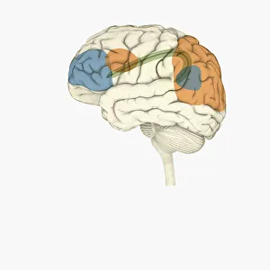
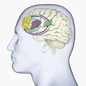
Male brain, computer artwork
men, cerebellum, cerebrum, spinal column, head and shoulders, side view, anatomy, illustration, black background, translucent, nobody, human body, brain stem, human representation, male likeness
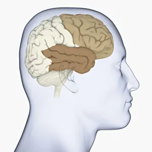
Male brain, computer artwork
men, cerebellum, cerebrum, spinal column, head and shoulders, side view, anatomy, illustration, black background, translucent, nobody, human body, brain stem, human representation, male likeness