mail_outline sales@mediastorehouse.com
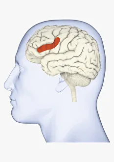
Digital illustration of head in profile showing area of mirror neuron in brain highlighted in orange
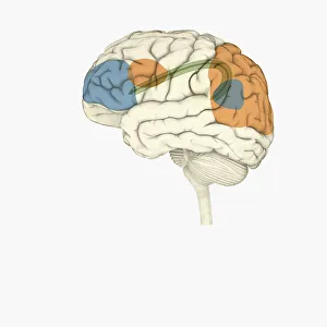
Digital illustration of frontal lobe and parietal lobe areas (orange) in left hemispheres (blue), and pathway of data from parietal lobe to frontal lobe (green) in human brain
Digital illustration of right superior temporal sulcas, and anterior cingulate cortex highlighted in red and grey in human brain
Digital illustration of anterior insular, anterior cingulate cortex, and ventromedial prefrontal cortex highlighted in human brain
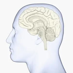
Digital illustration of head in profile showing brain
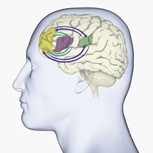
Digital illustration of head in profile showing memory areas of brain
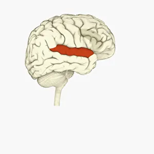
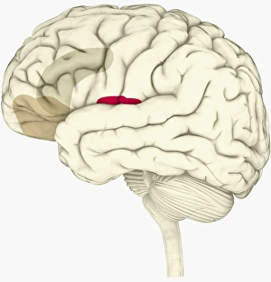
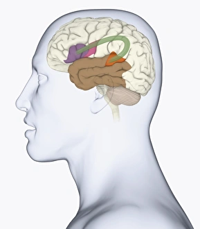
Digital illustration of head in profile showing bundle of nerve fibres connecti ng Brocas area and Wernickes area in human brain
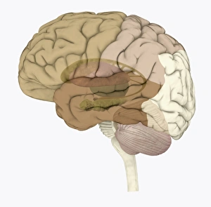
Digital illustration of areas associated with memory in human brain
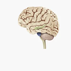
Digital illustration of hippocampus (green) and pons (blue) in left hemisphere of human brain
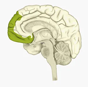
Digital illustration of human brain showing frontal cortex in green
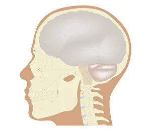
Cross section biomedical illustration of the brain and skull at 18 years of age