mail_outline sales@mediastorehouse.com
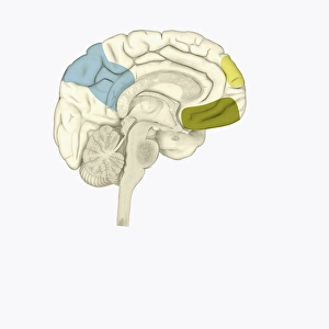
Digital illustration of posterior cingulate cortex (blue), medial frontal gyrus (yellow), and orbitofrontal prefrontal cortex (green) in human brain
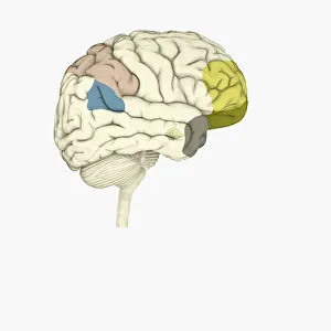
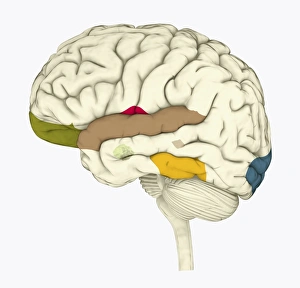
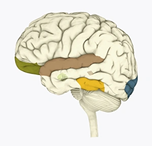
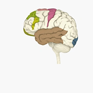
Digital illustration of parietal lobe (green), posterior superior temporal sulcas (blue), temperal pole (grey), dorsolateral prefrontal cortex, amygdala
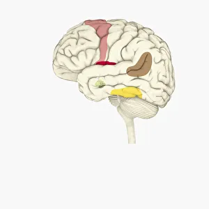
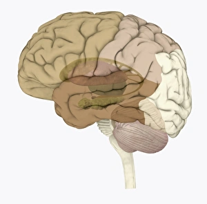
Digital illustration of emotional response areas in human brain
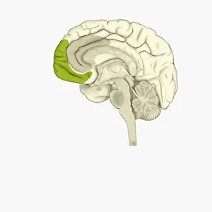
Digital illustration of anterior cingulate cortex (grey), and medial frontal cortex (green) in human brain
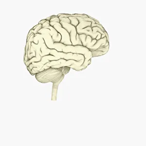
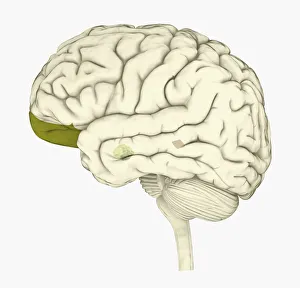
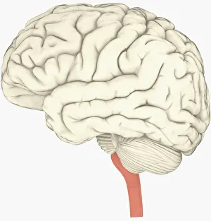
Digital illustration of human brain
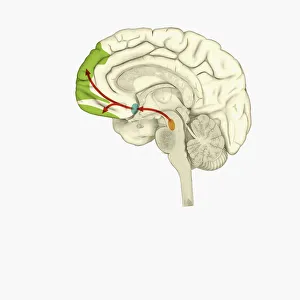
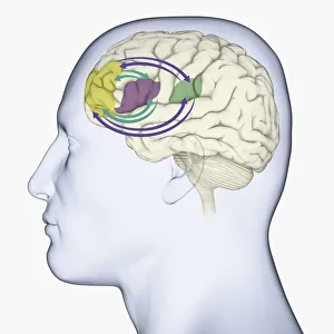
Digital illustration of reward pathway in human brain
Digital illustration of human brain associated with full awareness
Digital illustration of areas of information highlighted in human brain
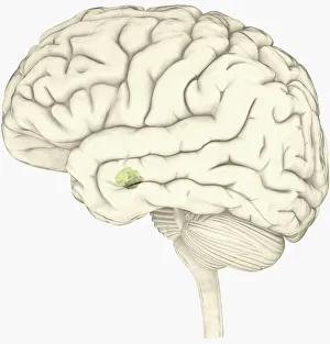
Digital illustration of human brain with orbitofrontal cortex and amygdala highlighted in green
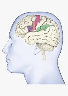
Digital illustration of head in profile showing mirror neurons in human brain highlighted in purple, pink and green
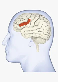
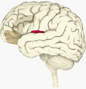
Digital illustration of head in profile showing area of mirror neuron in brain highlighted in orange
Digital illustration of prefrontal cortex of human brain highlighted in green
Digital illustration of various areas of cortex in human brain receiving input from sense organs
Digital illustration of crucial parts of highlighted in human brain
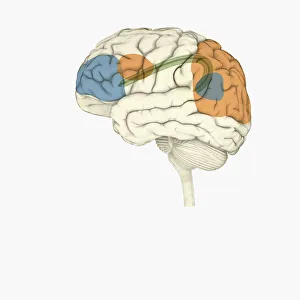
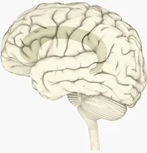
Digital illustration of frontal lobe and parietal lobe areas (orange) in left hemispheres (blue), and pathway of data from parietal lobe to frontal lobe (green) in human brain
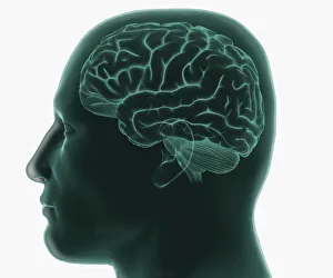
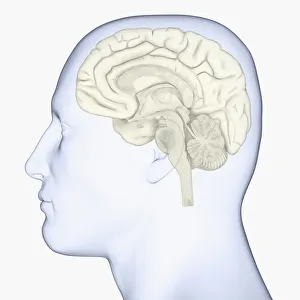
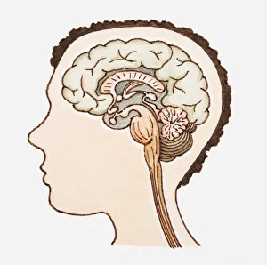
Digital illustration of human head in profile showing brain
Digital illustration of right superior temporal sulcas, and anterior cingulate cortex highlighted in red and grey in human brain
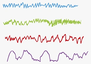
Digital illustration of alpha, beta, theta and delta brain waves
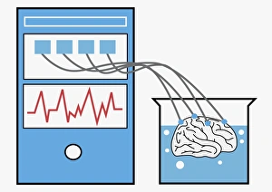
Digital illustration of computer providing stimulation to human brain, and brain experiencing virtual world
Digital illustration of anterior insular, anterior cingulate cortex, and ventromedial prefrontal cortex highlighted in human brain
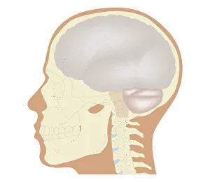
Digital illustration of head in profile showing skull, brain, and spine
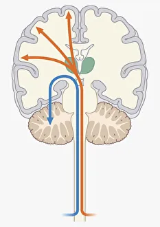
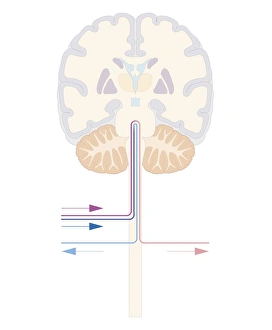
Digital illustration of unconscious and conscious pathways in human brain passing through thalamus ending in parietal lobe of cortex
Digital illustration of direction of dopamine flow, nucleus accumbens, basal ganglia and ventral tegmental area highlighted in human brain
Black and white digital illustration of cingulate cortex area of human brain
Digital illustration of amygdala in human brain
Digital illustration of human brain with spine highlighted in pink
Digital illustration of head in profile showing brain
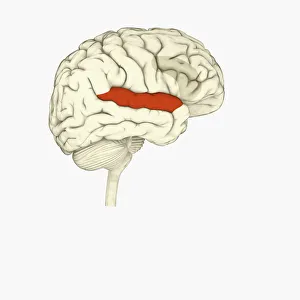
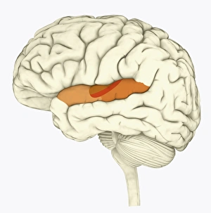
Digital illustration of human brain with primary auditory cortex highlighted in orange and red
Digital illustration of basal ganglia
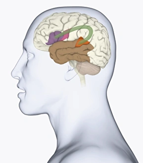
Digital illustration of head in profile showing memory areas of brain
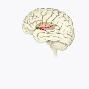
Digital illustration of head in profile showing bundle of nerve fibres connecti ng Brocas area and Wernickes area in human brain
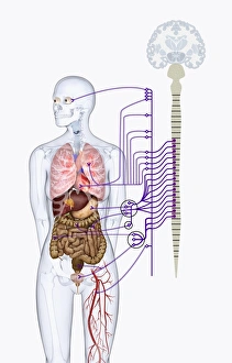
Digital illustration of autonomic nervous system responsible for automatic body functions
Digital illustration of areas associated with memory in human brain
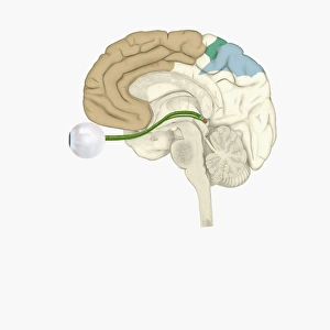
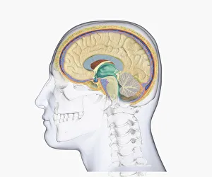
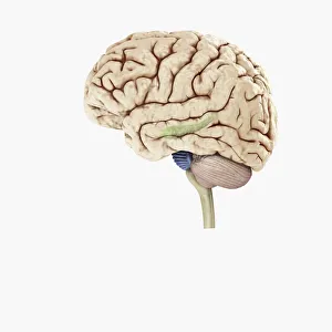
Digital illustration of hippocampus (green) and pons (blue) in left hemisphere of human brain
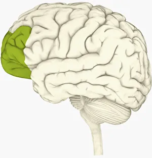
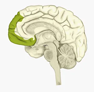
Digital illustration of human brain showing frontal cortex in green
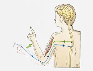
Illustration of reflex action in human arm
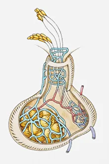
Illustration of pituitary gland releasing hormones
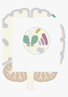
Digital illustration of human brain in yellow circle on white background
Illustration of human brain
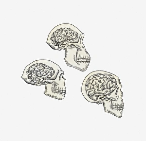
Illustration of skulls of Australopithecus, Homo erectus and Homo sapiens
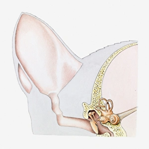
Cross section illustration of ear of domestic cat (Felis Catus)
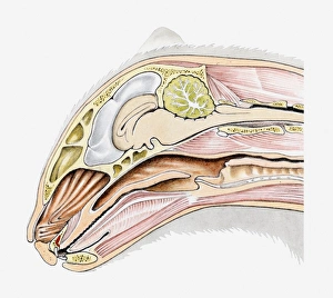
Cross section illustration of domestic cat (Felis Catus) head in profile
Cross section biomedical illustration of the brain and skull at 18 years of age
Cross section biomedical illustration of voluntary pathway of human brain
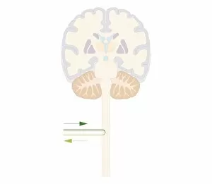
Cross section biomedical illustration of brain and autonomic pathways in the autonomic nervous system
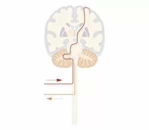
Cross section biomedical illustration of spinal reflex pathway of human brain
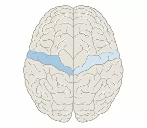
Cross section biomedical illustration of touch map of human brain
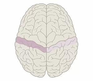
Cross section biomedical illustration of motor map of the brain