mail_outline sales@mediastorehouse.com
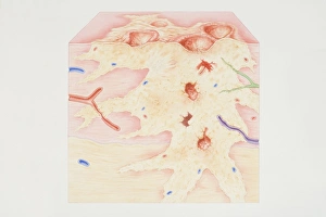
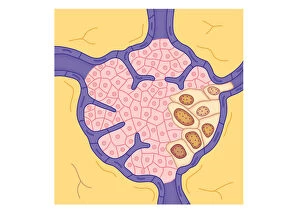
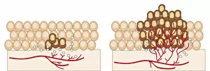
Cross-section diagram of a cancerous tumour including calcium deposits, blood vessels, tumour outgrowth, epithelial layer, ulcerated area, bleeding, nerve fibres, dead tissue and a lymph vessel
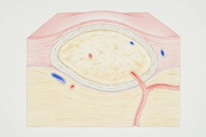
Cross-section diagram of a non-cancerous tumour including a fibrous capsule, tissue layer and blood vessel
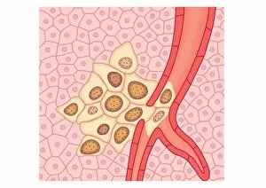
Digital illustration showing secondary tumour where cancerous cells have divided and lodged in narrow blood vessel and surrounding tissue
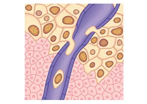
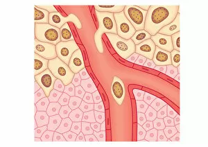
Digital illustration of breached lymph vessel as primary tumour grows and its cells invade adjacent tissues
Digital illustration of tumour in lymph node
Digital illustration of blood vessel wall rupturing, primary tumour expanding, cells rupturing walls of blood vessels, enabling cancerous cells to detach and spread via blood flow
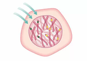
Digital illustration showing temporary cell damage to genes on chromosomes caused by an attacking carcinogen
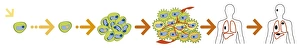
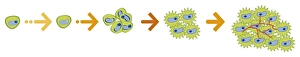
Digital illustration of the evolution of a cancer from initiation to metastasis
Digital illustration of angiogenesis process showing dormant tumour invading tissue, growing to mali
Digital illustration of action pathways of anti-cancer agents
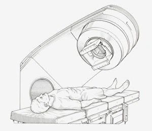
Black and white illustration of a patient undergoing radiation therapy