mail_outline sales@mediastorehouse.com
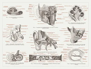
Anatomy of the human ear, lithograph, published in 1876Anatomy of the human ear. Lithograph, published in 1876
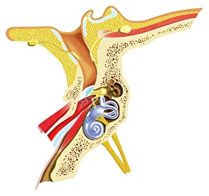
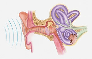
Diagram of inner ear showing auditory canal, eardrum, semicircular canals, cochlea, cochlea nerve, eustachian tube
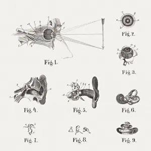
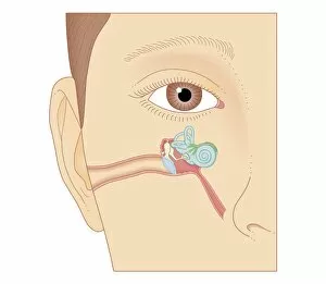
Anatomy of the human eye and ear, published in 1861Anatomy of the human eye and ear: 1) eye and eye socket, 2) eyeball (cross section, 3) eyeball with open sclera, 4) ear canals (cross section), 5) insulated inner hearing organs with earlobe
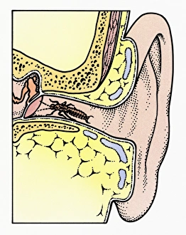
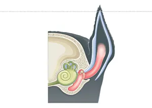
Cross section illustration of Common Earwig (Forficula auricularia) in auditory canal of ear, touching tympanic membrane with antennae
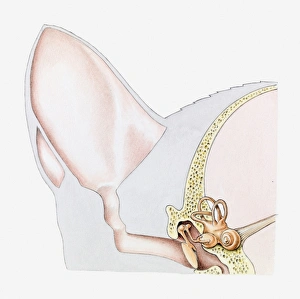
Digital cross section illustration of mammalian ear including pinna, ear drum, middle ear
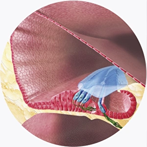
Digital illustration of Corti organ found in cochlea of human ear
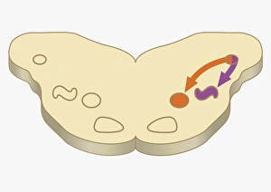
Digital cross section illustration of impulses passing from nearer cochlea nucleus to lateral superior olive
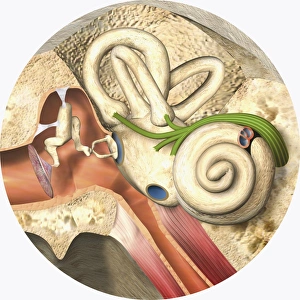
Digital illustration of middle and inner ear
Anatomical illustration of sound vibrations entering ear
Cross section illustration of ear of domestic cat (Felis Catus)
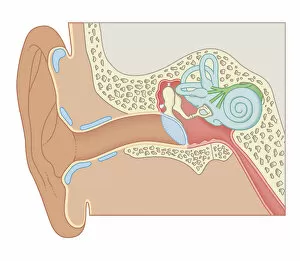
Cross section biomedical illustration of internal components of the ear
Cross section biomedical illustration of the anatomy of the ear
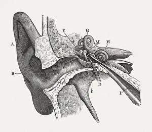
The human ear, wood engraving, published in 1880Anatomy of the human ear: A) auricle, B) External Auditory Canal, C) Tympanic Membrane, D) Tympanic Cavity, E) Malleus, M) Incus, H) Cochlea, G) Semicircular Canals, I) Eustachian Tube