mail_outline sales@mediastorehouse.com
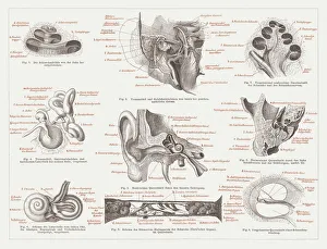
Anatomy of the human ear, lithograph, published in 1876Anatomy of the human ear. Lithograph, published in 1876
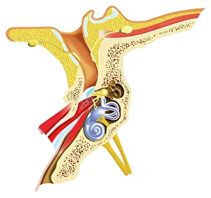
Diagram of inner ear showing auditory canal, eardrum, semicircular canals, cochlea, cochlea nerve, eustachian tube
Sweden. Uppsala Cathedral, the highest in ScandinaviaIllustration engraving of a Uppsala Cathedral, the highest in Scandinavia
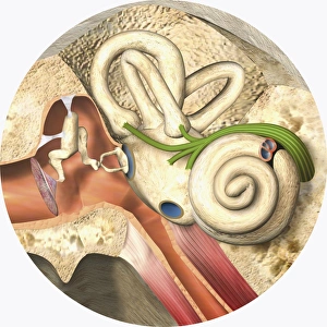
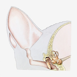
Digital cross section illustration of mammalian ear including pinna, ear drum, middle ear
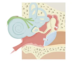
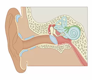
Digital illustration of middle and inner ear
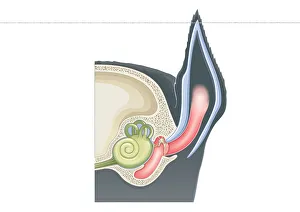
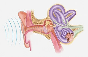
Anatomical illustration of sound vibrations entering ear
Cross section illustration of ear of domestic cat (Felis Catus)
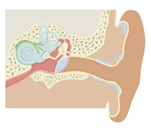
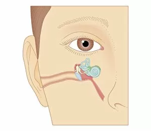
Cross section biomedical illustration of human ear
Cross section biomedical illustration of grommet inserted in eardrum (tympanic membrane)
Cross section biomedical illustration of internal components of the ear
Cross section biomedical illustration of the anatomy of the ear
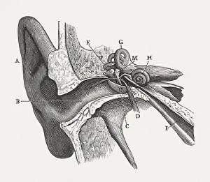
The human ear, wood engraving, published in 1880Anatomy of the human ear: A) auricle, B) External Auditory Canal, C) Tympanic Membrane, D) Tympanic Cavity, E) Malleus, M) Incus, H) Cochlea, G) Semicircular Canals, I) Eustachian Tube