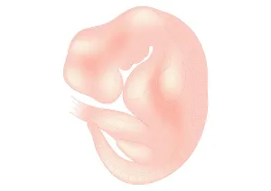
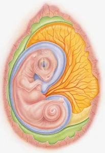
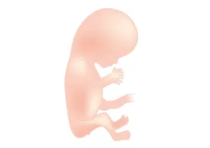
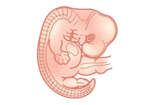
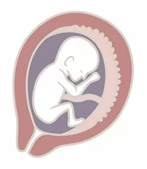
Digital illustration of foetus size at 6 weeks
Anatomy, Beginnings, Biomedical Illustration, Caucasian Appearance, Close-Up, Development, Digitally Generated, Foetus, Full Length, Growth, Healthcare and Medicine, Illustrative Technique, New Life
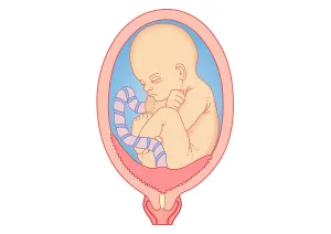
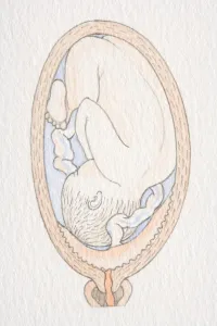
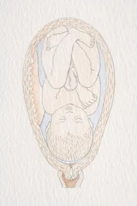
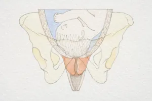
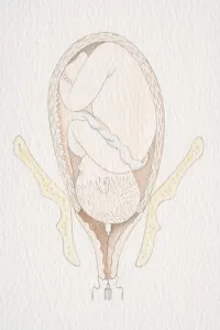
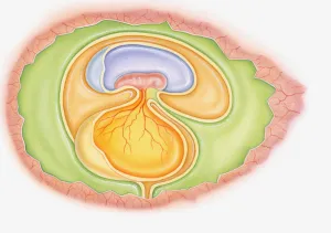
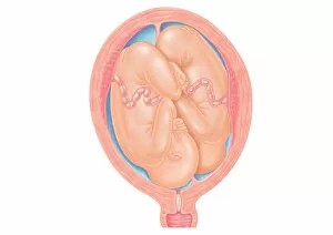
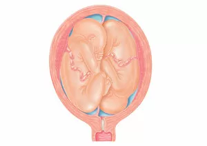
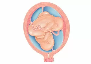
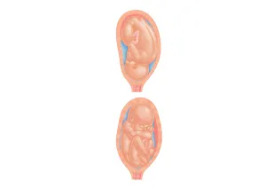
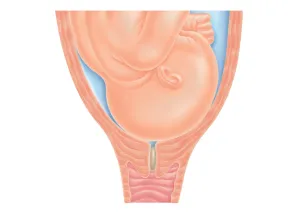
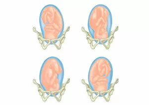
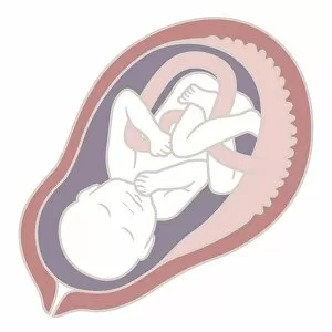
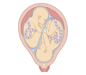
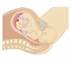
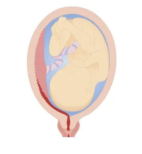
Digital illustration showing placenta praevia where the placenta is attached to the uterine wall
Anatomy, Baby, Beginnings, Biomedical Illustration, Blood Supply, Caucasian Appearance, Cervix, Cross Section, Danger, Development, Digitally Generated, Foetus, Full Length, Growth