mail_outline sales@mediastorehouse.com
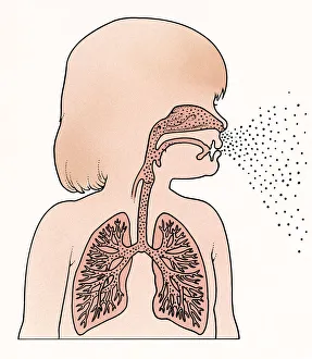
Cross section illustration of child in profile showing lungs and trachea, exhaling breath from mouth and nose
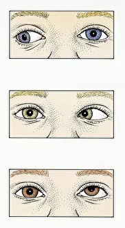
Sequence of illustrations showing Strabismus, Amblyopia, and Duane syndrome in children
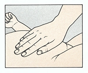
Illustration showing physical examination of babys abdomen to check for organ enlargement or abnormalities immediately after birth
Illustration of House Dust Mite (Dermatophagoides pteronyssinus)
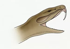
Illustration of snake showing needle-sharp, recurved tooth
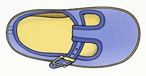
Illustration of childs comfortable shoe
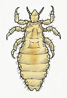
Illustration of dorsal view of male Body Louse (Pediculus humanus var. corporis)
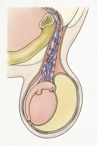
Cross section Illustration showing infantile hydrocele testis, with clear, amber-coloured fluid from spermatic cord surrounding testicle, resulting in swelling
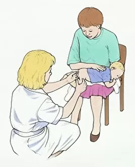
Illustration of nurse injecting vaccination in to leg of baby held by mother
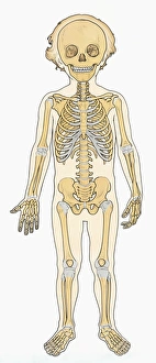
Illustration of skeleton of young boy
Digital illustration of biohazard symbol
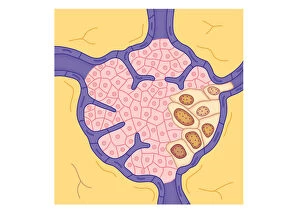
Digital illustration showing secondary tumour where cancerous cells have divided and lodged in narrow blood vessel and surrounding tissue
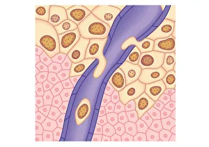
Digital illustration of breached lymph vessel as primary tumour grows and its cells invade adjacent tissues
Digital illustration of tumour in lymph node
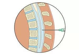
Digital illustration of lumbar puncture using spinal needle inserted into lumbar vertebrae and dura mater
Digital illustration of X-linked recessive inheritance
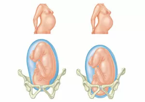
Digital illustration of pregnant two women and position of each babys head entering and inside pelvic area
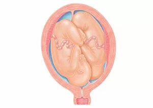
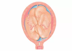
Cross section digital illustration of twins in normal position in uterus
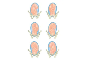
Six cross section digital illustrations of foetus showing position head in pelvis
Digital illustration showing baby breastfeeding
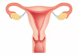
Digital cross section illustration of human uterus, fallopian tubes, ovaries, cervix, and vagina
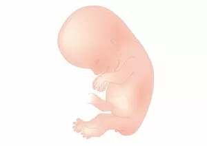
Digital illustration of foetal size at 11 weeks
Cross section digital illustration showing normal and breech positions
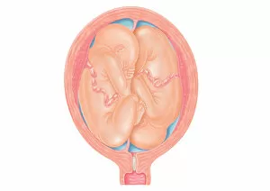
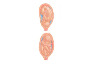
Cross section digital illustration of twins showing normal foetal presentation and transverse lie
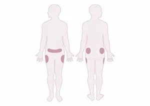
Digital illustration representing areas of fat distribution on belly, hips and thighs
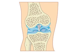
Digital cross section illustration of torn cartilage in knee
Cross section digital illustration of foetus in normal position and breech position
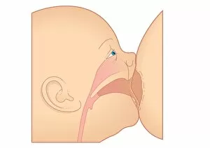
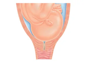
Cross section digital illustration of head of foetus in pelvis, pushing against cervix as labour nears, also showing mucus plug in cervix
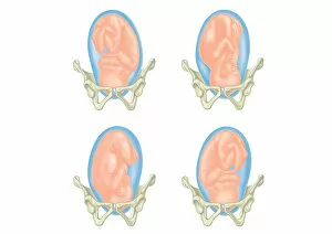
Four cross section digital illustrations showing head of foetus in pelvis
Digital illustration of foetus size at 7 weeks
Digital illustration of foetal size at 10 weeks
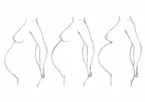
Digital illustration of bump position during pregnancy
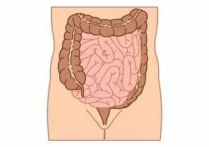
Digital illustration of location of intussusception in small intestine
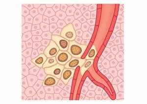
Digital illustration of blood vessel wall rupturing, primary tumour expanding, cells rupturing walls of blood vessels, enabling cancerous cells to detach and spread via blood flow
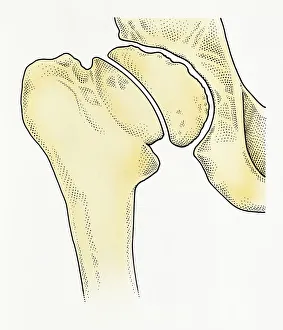
Digital illustration showing degeneration of hip joint known as Perthes disease
Digital illustration of recessive inheritance where both parents carry the abnormal Albinism gene
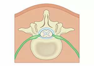
Digital illustration Spina Bifida, Meningocele, where protective covering around spinal cord protrudes through malformed vertebra to form sac filled with cerebrospinal fluid
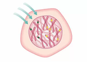
Digital illustration showing temporary cell damage to genes on chromosomes caused by an attacking carcinogen
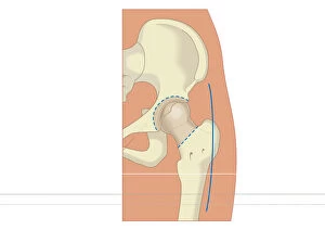
Digital illustration of prosthetic hip joint replacement
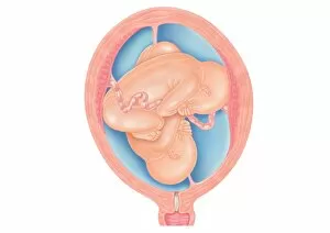
Cross section digital illustration of twins in breech position with buttocks presented first
Digital illustration DNA map
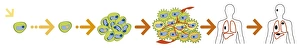
Digital illustration of the evolution of a cancer from initiation to metastasis
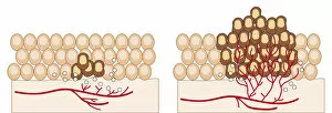
Digital illustration of angiogenesis process showing dormant tumour invading tissue, growing to mali