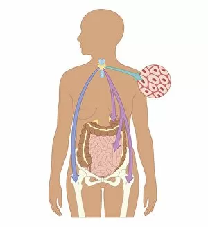
Human Internal Organ Collection (page 5)
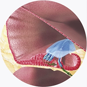
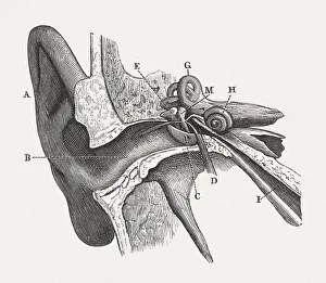
The human ear, wood engraving, published in 1880
Anatomy of the human ear: A) auricle, B) External Auditory Canal, C) Tympanic Membrane, D) Tympanic Cavity, E) Malleus, M) Incus, H) Cochlea, G) Semicircular Canals, I) Eustachian Tube
All Professionally Made to Order for Quick Shipping