mail_outline sales@mediastorehouse.com
Knee anatomy engraving 1866Atlas d anatomie descriptive du corps humain C. Bonamy - Paul Broca Victor Masson et Fils Paris 1866
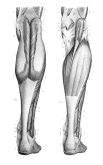
Posterior leg region anatomy engraving 1866Atlas d anatomie descriptive du corps humain C. Bonamy - Paul Broca Victor Masson et Fils Paris 1866
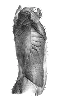
Side region of the trunk anatomy engraving 1866Atlas d anatomie descriptive du corps humain C. Bonamy - Paul Broca Victor Masson et Fils Paris 1866
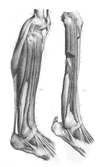
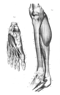
External leg region anatomy engraving 1866Atlas d anatomie descriptive du corps humain C. Bonamy - Paul Broca Victor Masson et Fils Paris 1866
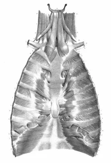
Sternum anatomy engraving 1866Atlas d anatomie descriptive du corps humain C. Bonamy - Paul Broca Victor Masson et Fils Paris 1866
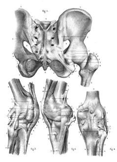
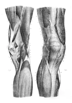
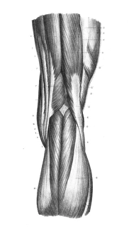
Anterior knee region anatomy engraving 1866Atlas d anatomie descriptive du corps humain C. Bonamy - Paul Broca Victor Masson et Fils Paris 1866
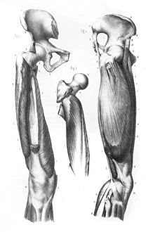
Femoral region anatomy engraving 1866Atlas d anatomie descriptive du corps humain C. Bonamy - Paul Broca Victor Masson et Fils Paris 1866
anatomy engraving 1866Atlas d anatomie descriptive du corps humain C. Bonamy - Paul Broca Victor Masson et Fils Paris 1866
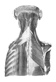
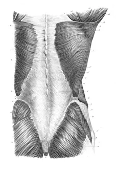
Back trunk anatomy engraving 1866Atlas d anatomie descriptive du corps humain C. Bonamy - Paul Broca Victor Masson et Fils Paris 1866
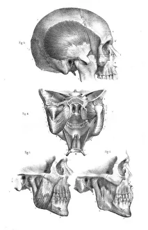
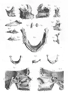
Skull mandibular anatomy engraving 1866Atlas d anatomie descriptive du corps humain C. Bonamy - Paul Broca Victor Masson et Fils Paris 1866
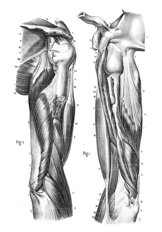
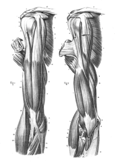
Region anterior of arm engraving 1866Atlas d anatomie descriptive du corps humain C. Bonamy - Paul Broca Victor Masson et Fils Paris 1866
Posterior part of knee engraving 1866Atlas d anatomie descriptive du corps humain C. Bonamy - Paul Broca Victor Masson et Fils Paris 1866
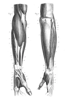
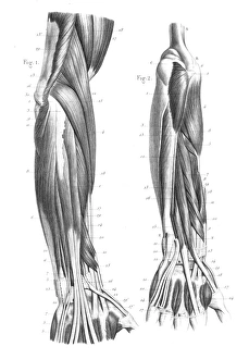
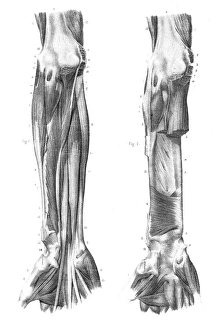
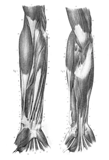
Posterior part of forearm engraving 1866Atlas d anatomie descriptive du corps humain C. Bonamy - Paul Broca Victor Masson et Fils Paris 1866
Region anterior of forearm engraving 1866Atlas d anatomie descriptive du corps humain C. Bonamy - Paul Broca Victor Masson et Fils Paris 1866
Face bones anatomy engraving 1866Atlas d anatomie descriptive du corps humain C. Bonamy - Paul Broca Victor Masson et Fils Paris 1866
Internal leg region anatomy engraving 1866Atlas d anatomie descriptive du corps humain C. Bonamy - Paul Broca Victor Masson et Fils Paris 1866
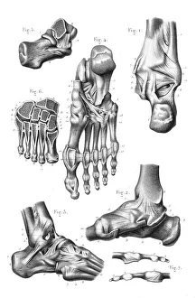
Foot joint anatomy engraving 1866Atlas d anatomie descriptive du corps humain C. Bonamy - Paul Broca Victor Masson et Fils Paris 1866
Anatomy arm engraving 1866Atlas d anatomie descriptive du corps humain C. Bonamy - Paul Broca Victor Masson et Fils Paris 1866
Region external of arm engraving 1866Atlas d anatomie descriptive du corps humain C. Bonamy - Paul Broca Victor Masson et Fils Paris 1866
Abdominal limbs anatomy engraving 1866Atlas d anatomie descriptive du corps humain C. Bonamy - Paul Broca Victor Masson et Fils Paris 1866
Back torso anatomy engraving 1866Atlas d anatomie descriptive du corps humain C. Bonamy - Paul Broca Victor Masson et Fils Paris 1866
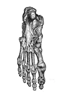
Foot bones and ligamentsAntique illustration of a foot bones and ligaments
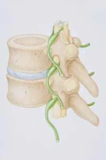
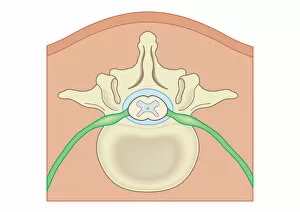
Section diagram depicting the human vertebral column, spinal nerve, spinal cord and vertebra of the delicate spina
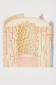
Cross-section diagram of a human long bone
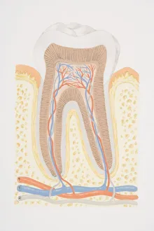
Cross-section diagram of tooth
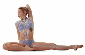
Woman sat with leg outstretched to one side, other leg folded in, arm behind head, skeleton, lymphatic system illustration overlay
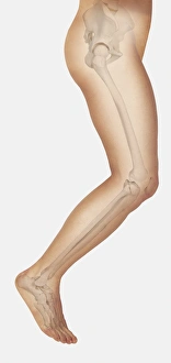
Diagram showing bones inside human leg, leaping forward
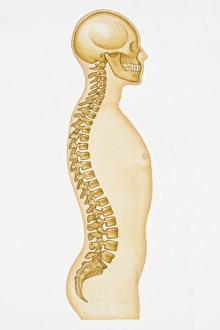
Illustration showing human spine and skull
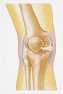
Illustration showing torn human knee ligament
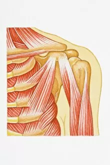
Illustration of separated shoulder, also known as a sprain, with circle around glenohumeral ligament
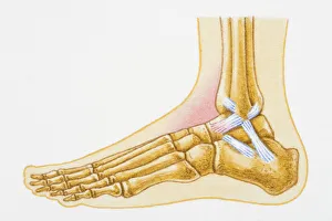
Illustration showing swollen talocrural joint caused by sprain to anterior talofibular ligament
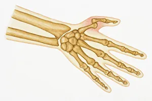
Illustration of bones in human hand and dislocated thumb
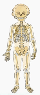
Illustration of skeleton of young boy
Digital illustration Spina Bifida, Meningocele, where protective covering around spinal cord protrudes through malformed vertebra to form sac filled with cerebrospinal fluid
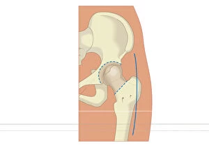
Digital illustration of prosthetic hip joint replacement
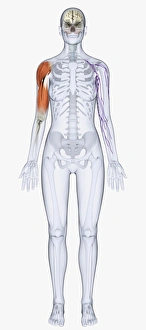
Digital illustration of human skeleton showing upper arm muscles
Sequence of illustrations showing complete bone fracture and healing process
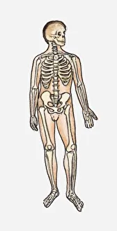
Illustration of skeletal system of the human body
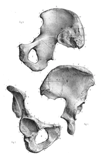
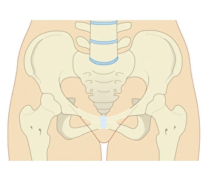
Cross section biomedical illustration of female type pelvis
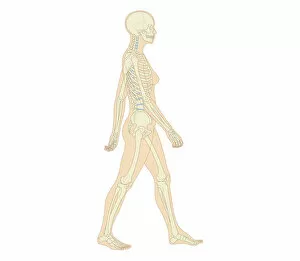
Cross section biomedical illustration of adult female human skeletal system and joints
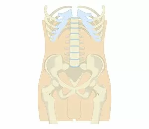
Cross section biomedical illustration of rib cage, spine and pelvis, and femur of adult male
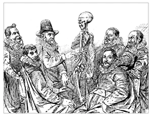
Antique illustration of 17th century anatomy lesson with skeleton: Doctor Egberts teach his students the skeleton structure (from a painting by the 17th century Dutch painter Thomas de Keyser)
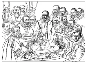
Antique illustration of 17th century anatomy lesson: doctor Van Der Meer dissect the body of a dead man and his students are around him at the university of Delft