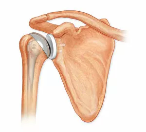
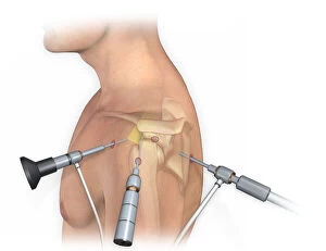
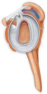
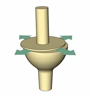
Anterior view total shoulder joint repair
Horizontal, Detail, Anatomy, Diagram, Repair, Illustration, Bone, Shoulder, Biology, Joint, Cut Out, White Background, Artwork, Color Image, Front View, Close Up, Healthcare And Medicine
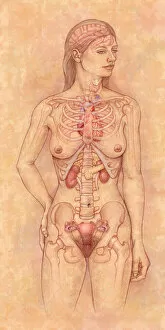
Front view of the female anatomy hilighting the endocrine system
Brain, Anatomy, Diagram, Heart, Illustration, Bone, Shoulder, Medical, Uterus, Biology, Pelvis, Femur, See Through, Vertical, Artwork, Female Likeness, Color Image, Front View
Anterior view of pelvis with hip bones showing arthritis and osteophytes on femoral heads
Horizontal, Anatomy, Diagram, Illustration, Bone, Disease, Medical, Hip, Biology, Pelvis, Femur, Cut Out, Arthritis, White Background, Artwork, Color Image, Front View, Healthcare And Medicine
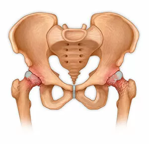
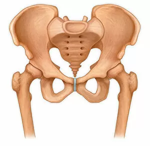
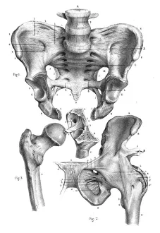
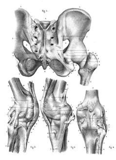
Normal anterior view of pelvis with hip bones
Horizontal, Anatomy, Diagram, Illustration, Bone, Medical, Hip, Biology, Pelvis, Femur, Cut Out, White Background, Artwork, Color Image, Front View, Healthcare And Medicine, The Human Body
Bovie used to cut through retincaculum, and clean up femur of Displaced patellar knee
Detail, Anatomy, Diagram, Muscle, Illustration, Bone, Cutting, Equipment, Injury, Medical, Tool, Biology, Femur, Surgery, Knee, Torn, Fractured, Meniscus, Cut Out, White Background, Vertical
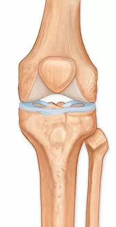
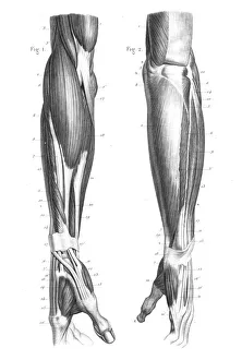
Illustration of the anterior knee, articular surface meniscus
anatomy, body part, bone, cartilage, close-up, color image, femur, fibula, front view, human body part, human bone, human joint, human knee, illustration, joint - body part, knee, patella, meniscus
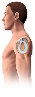
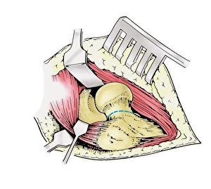
Lateral view of arthroscopic surgical repair on the shoulder joint
Horizontal, Detail, Anatomy, Diagram, Repair, Illustration, Bone, Shoulder, Equipment, Medical, Tool, Biology, Surgery, Joint, See Through, Cut Out, White Background, Artwork, Male Likeness
Open shoulder joint showing inflamed bursa from a bone spur and torn labrum
Detail, Anatomy, Diagram, Illustration, Bone, Shoulder, Injury, Medical, Biology, Joint, Torn, Injured, Cut Out, White Background, Vertical, Artwork, Color Image, Close Up, Healthcare And Medicine
Lateral view of the shoulder joint showing a tear in the labrum and an inflamed bursa
Anatomy, Diagram, Illustration, Shoulder, Injury, Medical, Biology, Surgery, Joint, See Through, Cut Out, White Background, Vertical, Artwork, Male Likeness, Color Image, Healthcare And Medicine
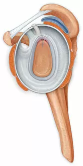
Lateral view of the shoulder joint showing the repair to the torn labrum
Anatomy, Diagram, Repair, Illustration, Shoulder, Injury, Medical, Biology, Surgery, Joint, Torn, See Through, Cut Out, White Background, Vertical, Artwork, Male Likeness, Color Image
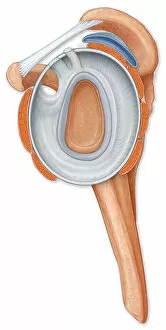
Lateral view of the shoulder joint showing the repair to the labrum
Detail, Anatomy, Diagram, Muscle, Illustration, Bone, Shoulder, Medical, Biology, Joint, Cut Out, White Background, Vertical, Artwork, Color Image, Close Up, Healthcare And Medicine, The Human Body
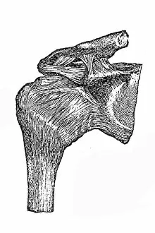
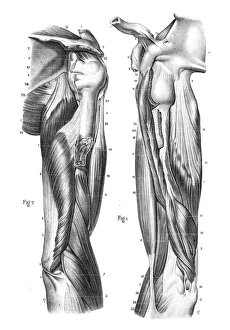
Normal side view of the shoulder joint hilighting the labrum, coracocromial ligament
Detail, Anatomy, Diagram, Muscle, Illustration, Shoulder, Medical, Biology, Joint, Side View, Cut Out, White Background, Vertical, Artwork, Color Image, Close Up, Healthcare And Medicine
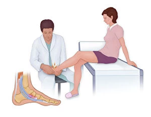
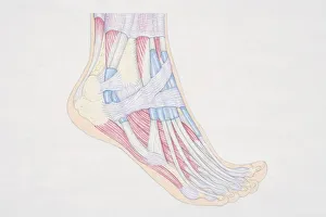
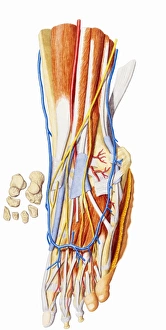
Accessory Navicular Pain Syndrome is caused by friction
Horizontal, Anatomy, Diagram, Illustration, Foot, Bone, Pressure, Injury, Medical, Doctor, Physician, Patient, Biology, Joint, Side View, See Through, Cut Out, White Background, Artwork
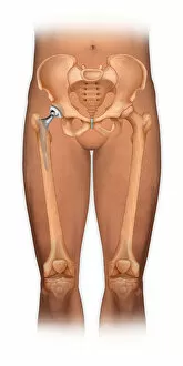
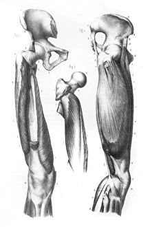
Front view of a body showing a total hip replacement
Detail, Anatomy, Diagram, Illustration, Bone, Disease, Medical, Hip, Biology, Pelvis, Femur, Acetabulum, Surgery, See Through, Cut Out, White Background, Vertical, Artwork, Color Image, Front View
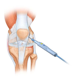
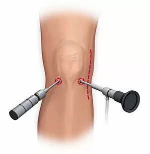
Arthroscopic surgical repair of the knee
Detail, Anatomy, Diagram, Repair, Illustration, Equipment, Medical, Tool, Biology, Surgery, Joint, Knee, Portals, Cut Out, White Background, Vertical, Artwork, Color Image, Front View, Close Up