mail_outline sales@mediastorehouse.com
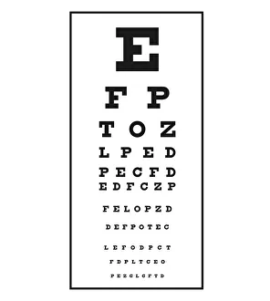
Black and white illustration of Snellen chart used to measure visual acuity. Snellen charts are named after the Dutch ophthalmologist Herman Snellen, who developed the chart in 1862
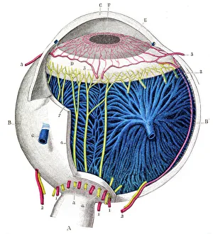
Human eye anatomy engraving 1899Corso Elementare di Scienze Naturali
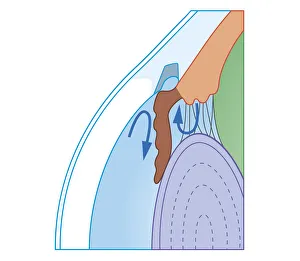
Cross section biomedical illustration of fluid flow in chronic glaucoma with blocked trabecular meshwork
Cross section biomedical illustration of Stereopsis
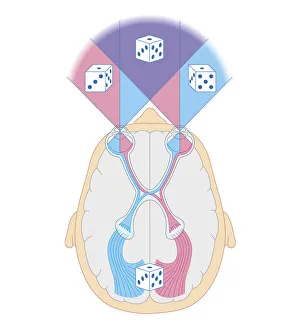
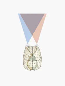
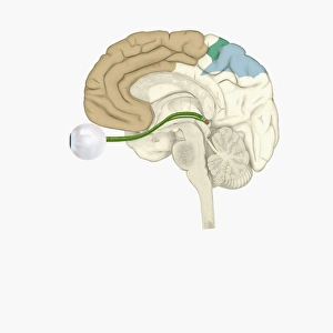
Digital illustration of human brain and information from left side of visual cortex receiving information from right visual field
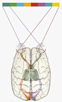
Digital illustration of human brain and mapping of visual field onto retina matching arrangement of data on surface of visual cortex
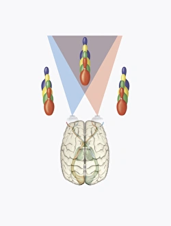
Digital illustration of human brain with differing views provided by each eye producing three dimensional vision
Cross section digital illustration of retina with futuristic retinal implant
Digital illustration of various areas of cortex in human brain receiving input from sense organs
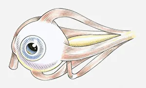
Illustration of muscles attached to human eye
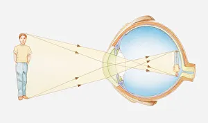
Illustration of vision of the human eye
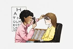
Illustration of optician doing an eye test on a patient
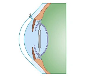
Cross section biomedical illustration of cataract surgery
Cross section biomedical illustration of laser surgery for retinopathy
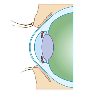
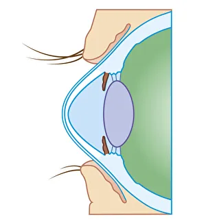
Cross section biomedical illustration of key anatomy of the eye
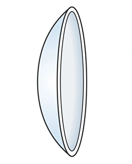
Illustration of hard contact lens
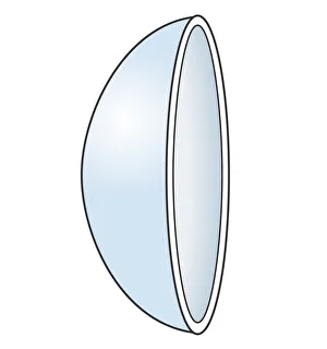
Illustration of soft contact lens
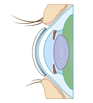
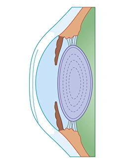
Cross section biomedical illustration of soft contact lens on eye
Cross section biomedical illustration of human eye before corrective surgery for myopia
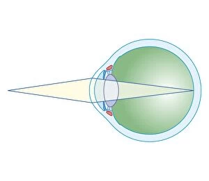
Cross section biomedical illustration of normal eye
Cross section biomedical illustration of keratoconus or conical cornea
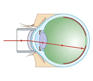
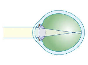
Cross section biomedical illustration of myopia
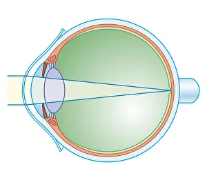
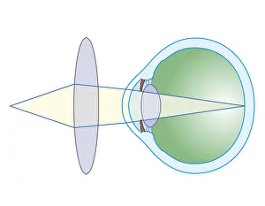
Cross section biomedical illustration of lens to correct hypermetropia
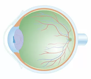
Cross section biomedical illustration of anatomy of human eye
Cross section biomedical illustration of eye focusing on near object