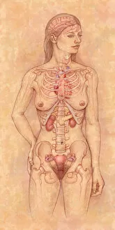
Front view of the female anatomy hilighting the endocrine system
Brain, Anatomy, Diagram, Heart, Illustration, Bone, Shoulder, Medical, Uterus, Biology, Pelvis, Femur, See Through, Vertical, Artwork, Female Likeness, Color Image, Front View
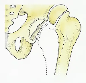
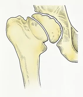
Digital illustration of dislocated hip
Anatomy, Biomedical Illustration, Close-Up, Cross Section, Digitally Generated, Dislocation, Healthcare and Medicine, Hip Bone, Human Bone, Illustration and Painting, Illustrative Technique
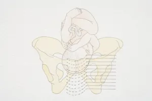
Anterior view of pelvis with hip bones showing arthritis and osteophytes on femoral heads
Horizontal, Anatomy, Diagram, Illustration, Bone, Disease, Medical, Hip, Biology, Pelvis, Femur, Cut Out, Arthritis, White Background, Artwork, Color Image, Front View, Healthcare And Medicine
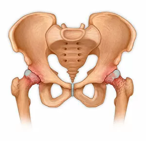
Normal anterior view of pelvis with hip bones
Horizontal, Anatomy, Diagram, Illustration, Bone, Medical, Hip, Biology, Pelvis, Femur, Cut Out, White Background, Artwork, Color Image, Front View, Healthcare And Medicine, The Human Body
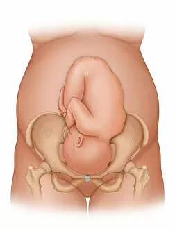
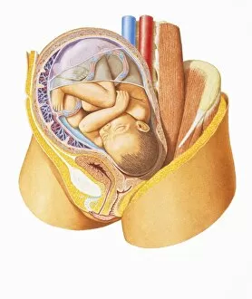
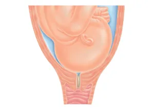
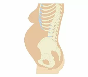
Front view of a woman nine months pregnant (baby phantomed within) ready for delivery
Detail, Baby, Anatomy, Diagram, Mother, Illustration, Bone, Birth, Medical, Fetus, Pregnant, Pregnancy, Biology, Pelvis, Femur, Development, Abdomen, Anterior, See Through, Cut Out, White Background
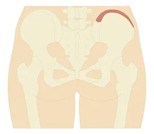
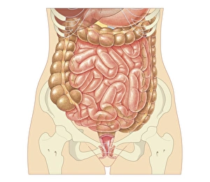
Repetitive activities like aerobics that involve lifting up the knee can cause trauma to
Detail, Anatomy, Diagram, Muscle, Pain, Illustration, Bone, Trauma, Injury, Medical, Biology, Pelvis, Femur, See Through, Cut Out, White Background, Artwork, Color Image, Front View, Close Up
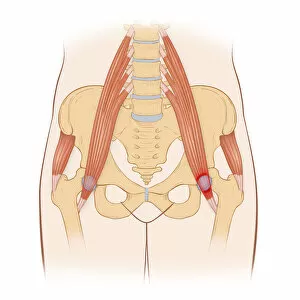
Snapping hip syndrome occurs when the iliopsoas tendon subluxes over the greater
Horizontal, Anatomy, Diagram, Muscle, Illustration, Bone, Medical, Hip, Biology, Pelvis, Femur, See Through, Rear View, Cut Out, Arthritis, White Background, Artwork, Color Image, Front View
Normal anatomy of an open hip showing the articular surface of the femur
Detail, Anatomy, Diagram, Illustration, Bone, Surface, Medical, Hip, Biology, Pelvis, Femur, Cut Out, White Background, Vertical, Artwork, Color Image, Close Up, Healthcare And Medicine
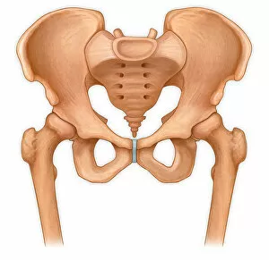
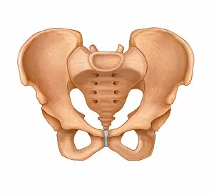
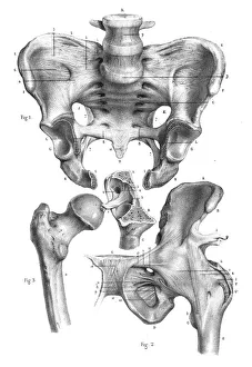
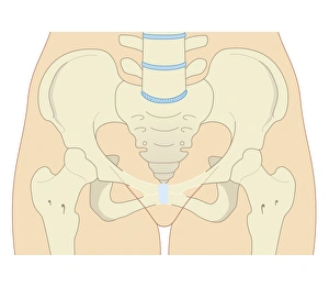
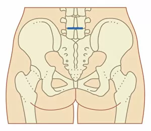
Normal anterior view of a pelvis
Horizontal, Anatomy, Diagram, Illustration, Bone, Medical, Hip, Biology, Pelvis, Cut Out, White Background, Artwork, Color Image, Front View, Healthcare And Medicine, The Human Body, No People
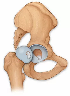
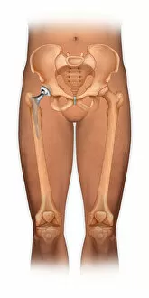
Front view of a body showing a total hip replacement
Detail, Anatomy, Diagram, Illustration, Bone, Disease, Medical, Hip, Biology, Pelvis, Femur, Acetabulum, Surgery, See Through, Cut Out, White Background, Vertical, Artwork, Color Image, Front View
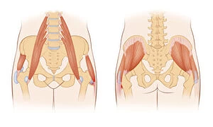
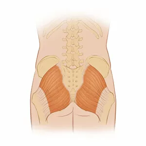
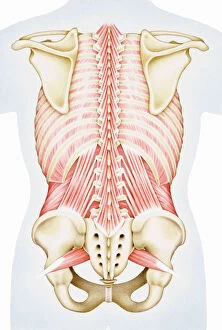
Normal posterior view of the hip bones and gluteus maximus muscle including the lumbar
Detail, Anatomy, Diagram, Muscle, Illustration, Bone, Medical, Hip, Biology, Pelvis, Femur, See Through, Rear View, Cut Out, White Background, Artwork, Color Image, Close Up, Healthcare And Medicine
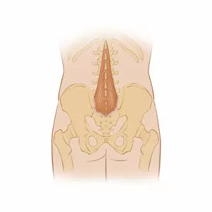
Normal posterior view of the back highlighting the multifidus muscle
Detail, Anatomy, Diagram, Muscle, Illustration, Ribs, Bone, Medical, Biology, Pelvis, See Through, Rear View, Cut Out, White Background, Artwork, Color Image, Close Up, Healthcare And Medicine