mail_outline sales@mediastorehouse.com
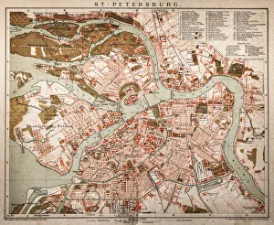
St. PetersburgAntique map of St. Petersburg
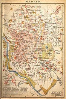
Map of MadridAntique map of Madrid from 1898
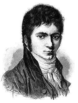
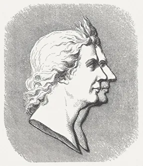
Antique illustration of young Beethoven
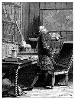
Antique illustration of scientific discoveries, electricity and magnetism: Benjamin Franklin
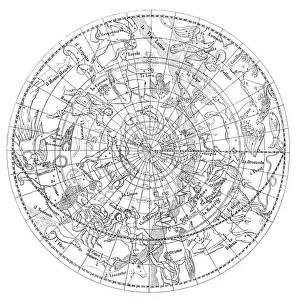
Antique illustration of astronomy constellations
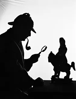
Silhouette of man wearing deerstalker, dressed as Sherlock Holmes. (Photo by HUNITED STATES - CIRCA 1950s: Silhouette of man wearing deerstalker, dressed as Sherlock Holmes
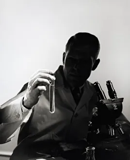
Male scientist wearing lab coat, sitting in darkness behind microscopeUNITED STATES - CIRCA 1950s: Male scientist wearing lab coat, sitting in darkness behind microscope, holding out test tube in light
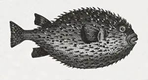
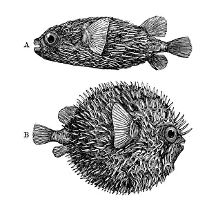
Blowfish EngravingEngraved illustrations of Members of Various Chordate Classes from Iconographic Encyclopedia of Science, Literature and Art, Published in 1851. Copyright has expired on this artwork
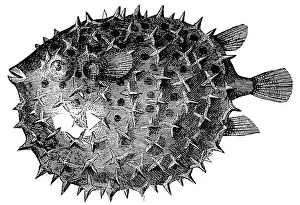
Antique illustration of Globe-fish (Diodon maculatus)
Long-spine Porcupinefish or Spiny Balloonfish (Diodon Holocanthus)
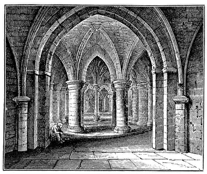
Antique illustration of Crypt, Canterbury Cathedral
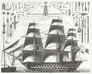
Engraving: French FrigateEngraved illustrations of a French Ship of the Line and Equipment from Iconographic Encyclopedia of Science, Literature and Art, Published in 1851. Copyright has expired on this artwork
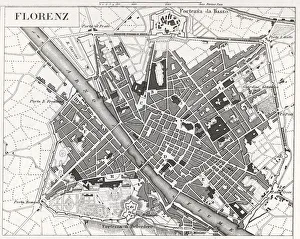
engraved illustrations of the city of FlorenceEngraved illustrations of the City of Florence from Iconographic Encyclopedia of Science, Literature and Art, Published in 1851. Copyright has expired on this artwork. Digitally restored
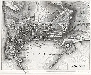
Engraving: Ancona, ItalyEngraved illustrations of the City of Ancona, Italy from Iconographic Encyclopedia of Science, Literature and Art, Published in 1851. Copyright has expired on this artwork. Digitally restored
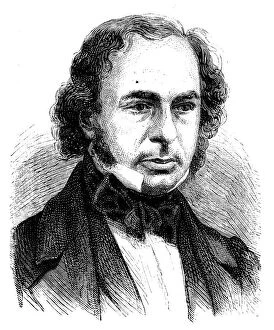
Antique illustration of scientist: Isambard Kingdom Brunel
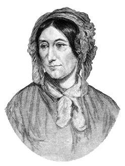
Mary Somerville, mathematician (1875 illustration)An illustration from " The Family Friend" published by S.W. Partridge & Co. (London, 1875). Mary Fairfax Greig Somerville, mathematician, scientist and astronomer
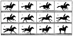
Antique illustration of horse in motion
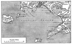
Antique map of Gulf of Naples
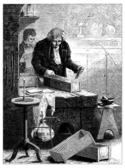
Antique illustration of scientific discoveries, electricity and magnetism: Cruikshank
The Brothers Montgolfier, inventors of the hot air balloonThe brothers Joseph Michel Montgolfier (1740 - 1810) and Jacques Etienne Montgolfier (1745 - 1799) was the inventors of the hot air balloon, the Montgolfiere (1783)
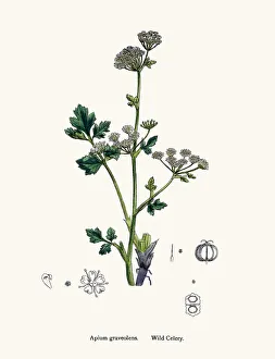
Wild celery plant used as diuretic and lazativeDigitally restored image of an original antique illustration by Sowerby published in 1860s in The English Botany
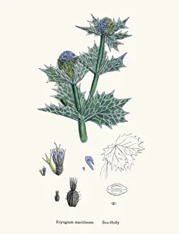
Sea Holly plant against bladder disease and as tonicDigitally restored image of an original antique illustration by Sowerby published in 1860s in The English Botany
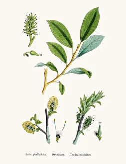
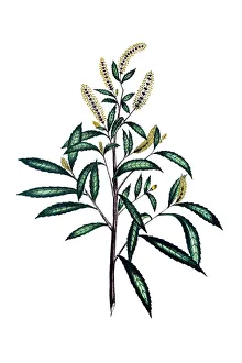
Willow medicinal tree remedy for aches and feverWillows, also called sallows, and osiers, form the genus Salix, around 400 species of deciduous trees and shrubs, found primarily on moist soils in cold
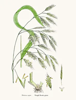
Brome grasses evasive weedsDigitally restored image of an original antique illustration by Sowerby published in 1860s in The English Botany
American dusky willow tree 19th century botanical. Digitally restored image of an original antique lithograph from North American Sylva book published in 1871
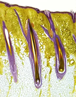
Human skin showing hair follicles, LMHairy skin. Light micrograph of a thick section of human skin, showing three central hair follicles. The outer layer of the skin, the epidermis, is the thin, purple band, supported by the deeper
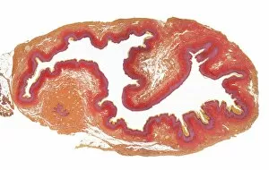
Cervix, LMCervix. Low power lght micrograph (LM) of the cervix. The cervix is the narrow inferior portion of the uterus. The part which projects into the vagina is seen here
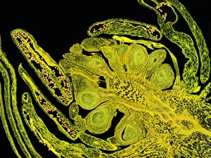
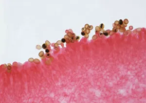
Flower bud, LMFlower bud. Light microscope image (LM) of a section of a flower bud. The small yellow pollen grains are visible in the anthers, the male part of the flower
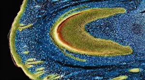
Developing nail, LMDeveloping nail. Light micrograph (LM) of longitudinal section through a fetal finger tip to show the developing nail. The large area of green-yellow nail bed epithelium is tipped by the developing
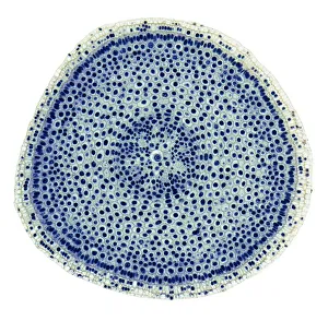
Onion root tip, LMMitosis. Light micrograph (LM) of a transverse section of onion (Allium cepa) root tip to show cells undergoing mitosis (nuclear division). Magnification: x100 when printed at 10 centimetres wide
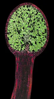
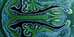
Liverwort spore capsule, LMLiverwort spore capsule. Light micrograph (LM). Longitudinal section through the thallus and sporangium of a liverwort (Pellia epiphylla)
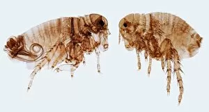
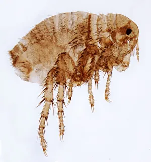
Human fleas, LMHuman fleas. Light micrograph (LM) of a male (left) and female human flea (Pulex irratans). Fleas are wingless and flattened from side to side, which makes them difficult to dislodge in hair
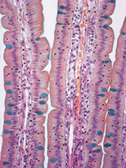
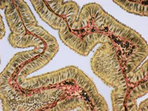
Small intestine, LMSmall intestine. Light micrograph (LM) of a section through the finger-like projections (villi) of the duodenum, the uppermost part of the small intestine
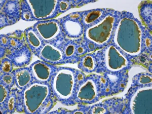
Thyroid, LMThyroid gland. Light micrograph (LM) of a thyroid gland showing the follicles. The follicles are lined by a single layer of cuboidal epithelial cells (blue)
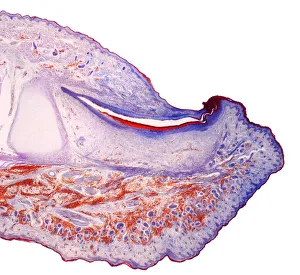
Fingertip, LMFingertip. Light micrograph (LM) of a section through the fingertip. The nail (orange) is at top center, with the nail root below. The nail bed is dark purple and is continous with the epithelium
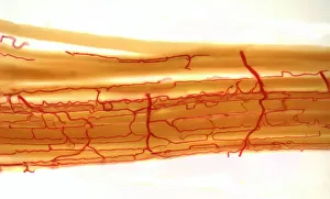
Blood supply to muscles, LMBlood supply to muscles. Light micrograph (LM) showing blood supply to muscle fibers. The muscle fibers (yellow) have been teased apart to reveal the capillary bed (red)
Fallopia tube, LMFallopian tube. Light micrograph (LM).The fallopian tube, or oviduct, conveys the egg from the ovary to the uterus. Ciliated columnar epithelium is yellow
Mushroom gill, LMMushroom gills. High power light micrograph (LM) of a section through the gills of a mushroom, Agaricus sp. (formerly Psalliota sp.)
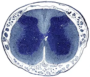
Spinal cord, LMSpinal cord. Light micrograph (LM) of a cross-section through the human spinal cord in the lumbar region. The spinal cord consists of a butterfly-shaped core (dark blue) known as grey matter
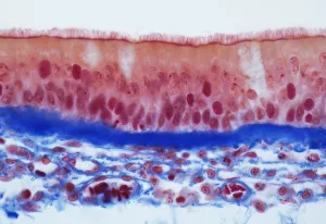
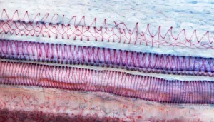
Trachel epithelium, LMTrachea epithelium. Light micrograph (LM) of a vertical section through the pseudostratified columnar epithelium from the trachea
Female flea, LMHuman flea. Light micrograph (LM) of a female human flea (Pulex irratans). Fleas are wingless and flattened from side to side, which makes them difficult to dislodge in hair
Nasal sinuses, LMNasal sinuses. Light micrograph (LM) of the nasal sinuses ( lined by cyan epithelium ) and the supporting cartilages (green). Bone tissue is identified by the blue bone marrow
Xylem, LMXylem tissue. Light micrograph (LM) of a section through sunflower(helianthus annuus) tissue showing spiral tracheids, a type of xylem