mail_outline sales@mediastorehouse.com
Illustration of Thomsons Plum Pudding model of the atom, with negatively charged electrons dotted
Illustration of two cross section blocks of earth divided by fault line
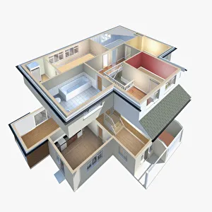
Model of a house with interior exposed
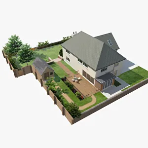
Model of a house with patio, garden and garden shed
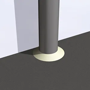
Downpipe connected directly to underground drainage, close-up
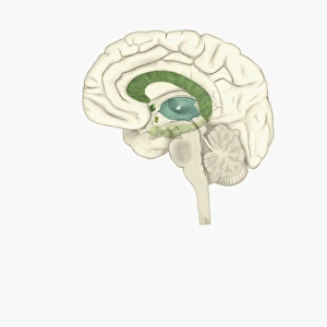
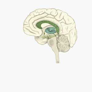
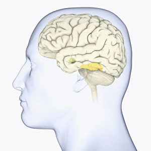
Digital illustration of striatum and amygdala highlighted in human brain
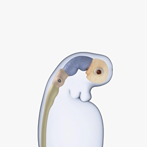
Digital illustration of neural tube, rudimentary eye and ear buds of three week old human embryo
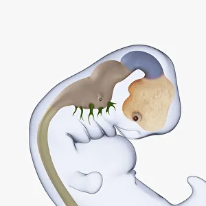
Digital illustration of 7 week old human embryo
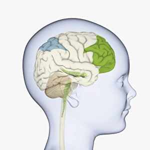
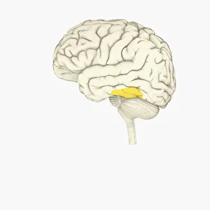
Digital illustration of childs head in profile highlighting parts of brain
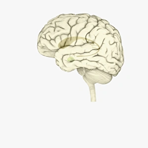
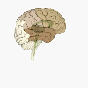
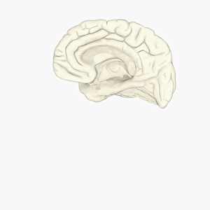
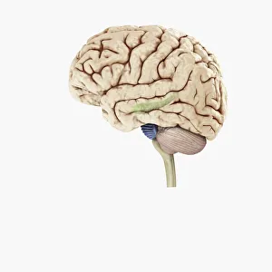
Digital illustration of showing left side of human brain
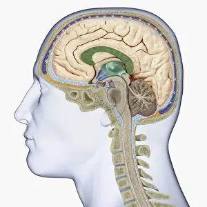
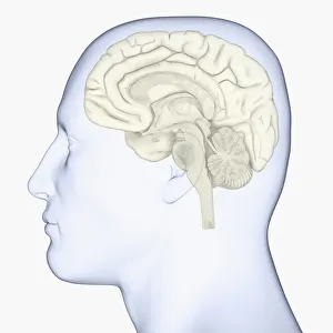
Digital illustration of head in profile showing cross section of brain, neck vertebra and spine
Digital illustration of showing right side human brain
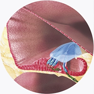
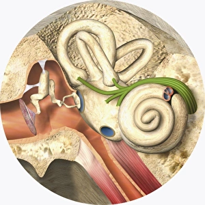
Digital illustration of Corti organ found in cochlea of human ear
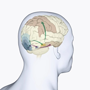
Digital illustration of head in profile showing dorsal and ventral pathways of brain
Cross section digital illustration of retina with futuristic retinal implant
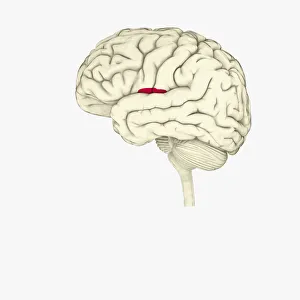
Digital illustration of human brain showing face-recognition area highlighted in yellow
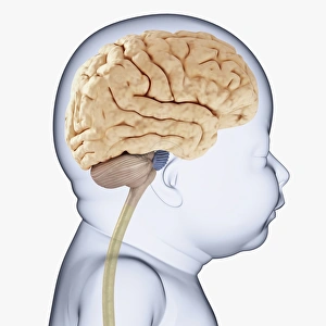
Digital illustration of head of baby in profile showing brain
Digital illustration of brain areas involved in altered states
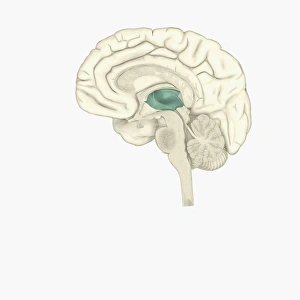
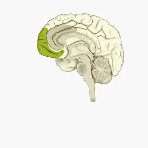
Digital illustration of thalamus in human brain highlighted in green
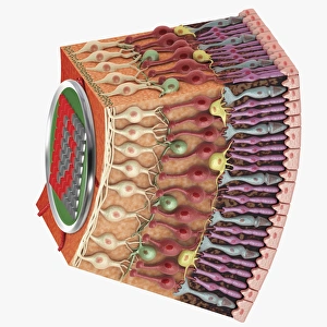
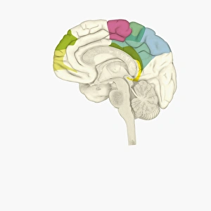
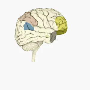
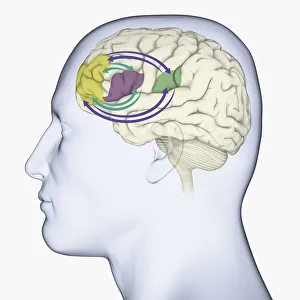
Digital illustration of parts of human brain highlighted in shades of green, pink, blue, and yellow
Digital illustration of 22 pairs of chromosomes plus one sex pair
Digital illustration of female human brain
Digital illustration of male human brain
Digital illustration of insula in human brain highlighted in red
Digital illustration of human brain showing corpus collosum and cingulate gyrus on medial surface of human brain
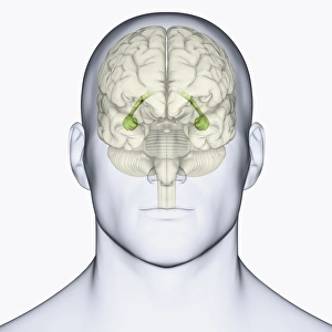
Digital illustration of head in profile showing face recognition area and amygdala in brain
Digital illustration of head showing location of hippocampus in human brain
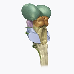
Digital illustration of human brain stem with cerebellum removed revealing medulla and axons
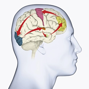
Digital illustration of head in profile showing motor cortex (pink), frontal area and amygdala (green), auditory cortex (orange), and visual cortex in brain
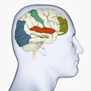
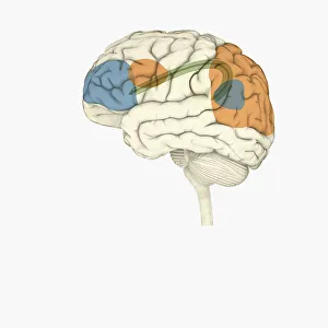
Digital illustration of head in profile showing visual cortex (blue), motor cortex (pink), auditory cortex (orange), frontal area and amygdala (green) brain
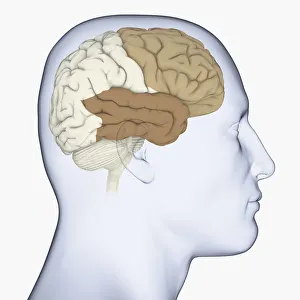
Digital illustration of head in profile showing frontal lobe and temporal lobe in brain
Digital illustration of posterior cingulate cortex (blue), medial frontal gyrus (yellow), and orbitofrontal prefrontal cortex (green) in human brain
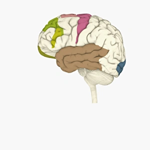
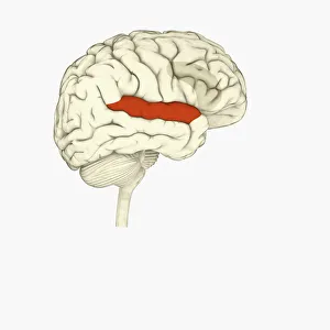
Digital illustration of parietal lobe (green), posterior superior temporal sulcas (blue), temperal pole (grey), dorsolateral prefrontal cortex, amygdala
Digital illustration of emotional response areas in human brain
Digital illustration of anterior cingulate cortex (grey), and medial frontal cortex (green) in human brain
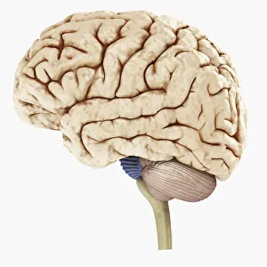
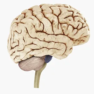
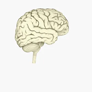
Digital illustration of human brain
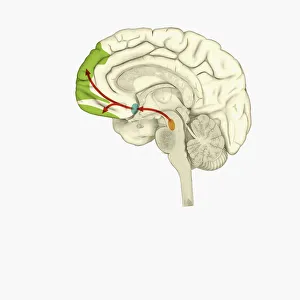
Digital illustration of reward pathway in human brain
Digital illustration of various areas of cortex in human brain receiving input from sense organs
Digital illustration of crucial parts of highlighted in human brain
Digital illustration of frontal lobe and parietal lobe areas (orange) in left hemispheres (blue), and pathway of data from parietal lobe to frontal lobe (green) in human brain
Digital illustration of right superior temporal sulcas, and anterior cingulate cortex highlighted in red and grey in human brain
Digital illustration of direction of dopamine flow, nucleus accumbens, basal ganglia and ventral tegmental area highlighted in human brain
Digital illustration of head in profile showing brain
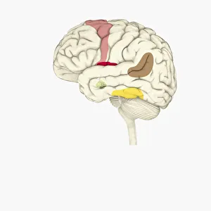
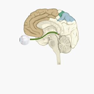
Digital illustration of head in profile showing memory areas of brain
Digital illustration of middle and inner ear
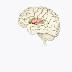
Digital illustration of hippocampus (green) and pons (blue) in left hemisphere of human brain
Digital illustration of white water droplet in blue circle on white background
Digital illustration of yoga lotus position in purple circle on white background