mail_outline sales@mediastorehouse.com
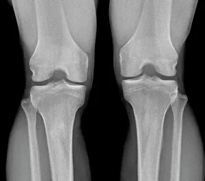
Normal knees, X-rayNormal knees. Frontal X-ray of the flexed knees of a 24 year old patient
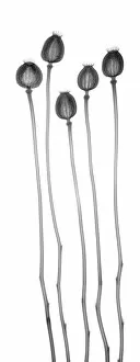
Poppy seed heads, X-ray
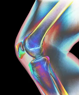
Normal knee, X-rayNormal knee. Coloured X-ray of the knee of a 44 year old woman
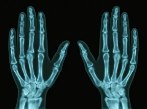
Hands, X-ray
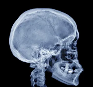
Normal skull, X-rayNormal skull. Coloured X-ray of the head of a 28 year old patient
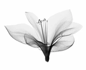
Amaryllis, X-ray
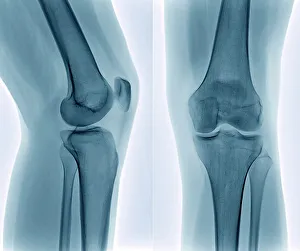
Healthy knee, X-rayHealthy knee. Coloured frontal (right) and profile (left) X-rays of the healthy knee of a 24 year old patient
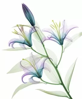
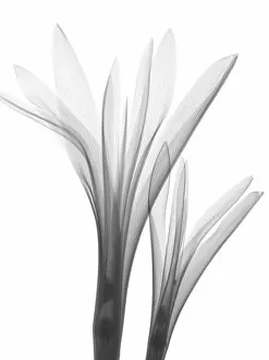
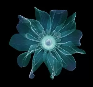
Oriental stargazer lily (Lilium sp. ), coloured X-rayOriental stargazer lily (Lilium sp.), coloured X-ray
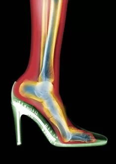
Leg in stiletto shoe MRI style, X-rayLeg in stiletto shoe, coloured MRI style X-ray
Parsley (Petroselinum crispum), X-ray
Tulip (Tulipa sp. ), X-rayTulip (Tulipa sp.), X-ray
Gladiolus, X-ray
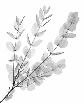
Gum tree (Eucalyptus cinerea), X-ray
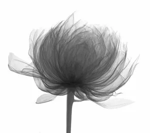
Peony head (Paeonia sp. ), X-rayPeony head (Paeonia sp.), X-ray
Red ginger flower and leaf, X-ray
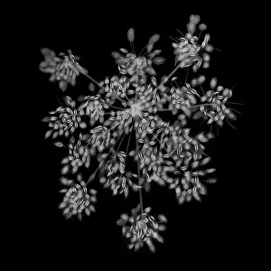
Allium flower, X-ray
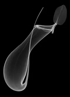
Pitcher plant (Nepenthes coccinea) pitcher, X-ray
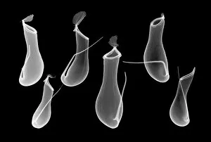
Six pitcher plant (Nepenthes coccinea) pitchers, X-ray
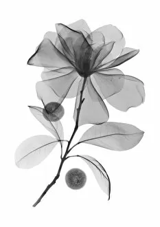
Magnolia (Magnolia virginiana) flower head, X-rayMagnolia (Magnolia virginiana) flower head, coloured X-ray
Great coneflower (Rudbeckia maxima), X-rayGreat coneflower (Rudbeckia maxima), coloured X-ray
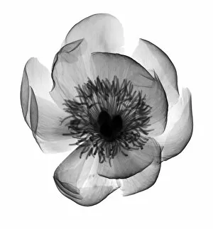
Hellebore (Helleborus hybridis), X-rayHellebore (Helleborus hybridis), coloured X-ray
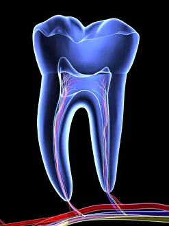
Molar toothTooth, transparent cross section of a molar tooth with arteries (red), veins (purple) and nerves (green)
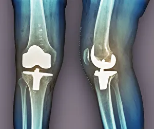
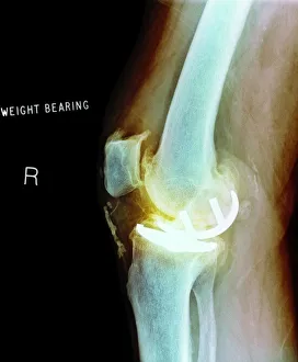
Total knee replacement, X-raysTotal knee replacement. Coloured frontal (left) and profile (right) X-rays of the right knee of a 69 year old patient after total knee replacement surgery
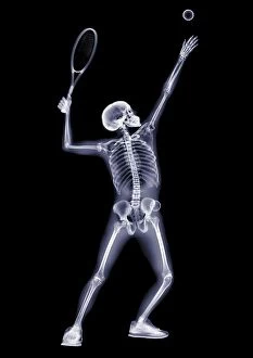
Person serving tennis ball, X-ray
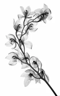
Cymbidium orchid, X-ray
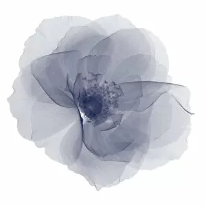
Magnolia flower, X-ray
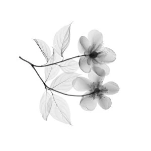
Orange blossom flowers, X-ray
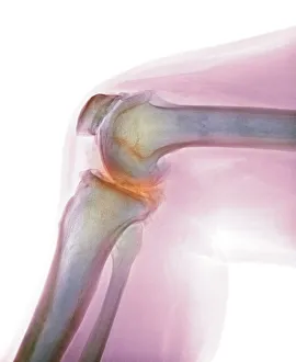
Arthritis of the knee, X-rayArthritis of the knee. Coloured X-ray the arthritic knee of a 37 year old man. The knee had previously been damaged in a motorbike accident
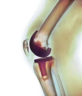
Knee replacement, X-rayKnee replacement. Coloured X-ray of a total knee replacement in a 70 year old man
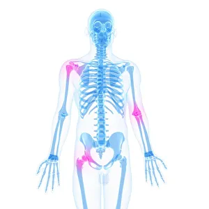
Joint pain, conceptual artworkJoint pain, conceptual computer artwork
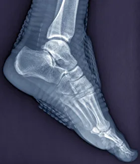
Healthy ankle joint, X-rayHealthy ankle joint. Coloured profile X-ray of the left ankle of a 21 year old patient. Strapping around the ankle is visible on this X-ray
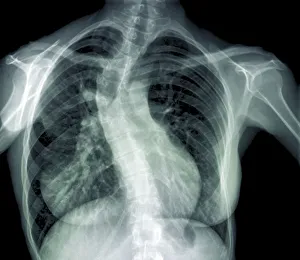
Scoliosis of the spine, X-rayScoliosis of the spine. Coloured X-ray of the chest of a 31 year old patient with scoliosis (sideways curvature) of the spine
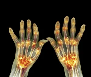
Rheumatoid arthritis, X-rayRheumatoid arthritis. Coloured X-ray of the hands of an 81 year old female patient with rheumatoid arthritis
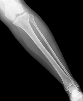
Normal lower leg, X-rayNormal lower leg. X-ray of the lower leg of a 24 year old woman
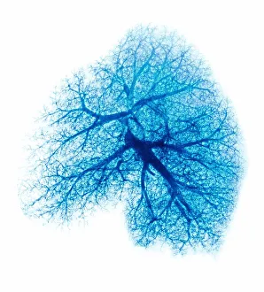
Lung, X-rayLung. Coloured X-ray showing the blood vessels in a lung
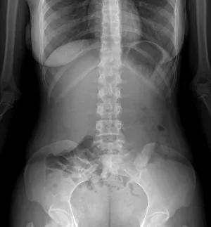
Normal abdomen, X-rayNormal abdomen. X-ray of the abdomen of a 20 year old female
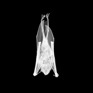
Flying fox, X-rayFlying fox, or fruit bat (Pteropus sp.), X-ray
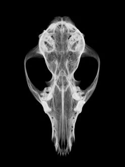
Fox skull, X-ray
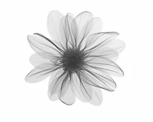
Michaelmas daisy (Aster amellus) flower head, X-ray
Peony (Paeonia sp. ), X-rayPeony (Paeonia sp.), X-ray
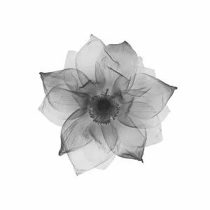
Lotus flower (Nelumbo nucifera), X-ray
Magnolia flower and acai berries, X-ray
Gardenia head from above, X-rayGardenia sp. head from above, X-ray
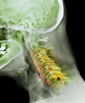
Arthritis of the neck, X-rayArthritis of the neck. Coloured X-ray of the arthritic cervical spine of a 70 year old man
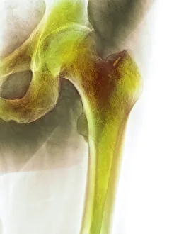
Normal hip, X-rayNormal hip. Coloured X-ray of the hip of a 90 year old man
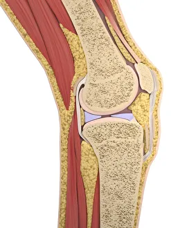
Knee anatomy, artworkKnee anatomy, computer artwork
Osteoarthritis of the knee, X-rayOsteoarthritis of the knee, coloured X-ray