mail_outline sales@mediastorehouse.com
1,262 items
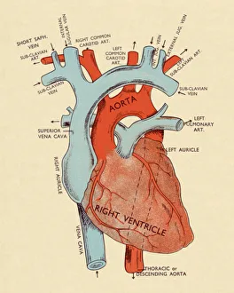
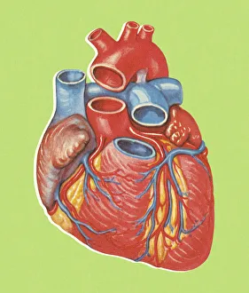
Diagram of Heart
Heart Diagram
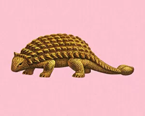
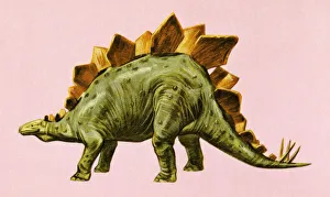
Dinosaur
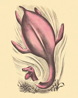
Dinosaur Swimming
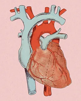
Human Heart
Stegosaurus Dinosaur
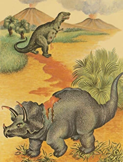
Dinosaur in the Wilderness
People Riding Dinosaurs
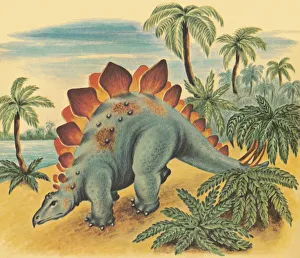
Dinosaur in the Jungle
Two Dinosaurs
Dinosaur Wading in a Pond
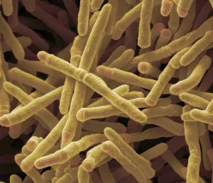
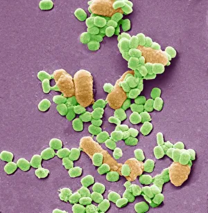
Mycobacterium smegmatis bacteria, SEMMycobacterium smegmatis bacteria. Coloured scanning electron micrograph (SEM) of Mycobacterium smegmatis bacteria. These bacteria feed on dead or decaying material
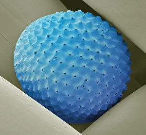
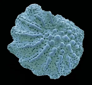
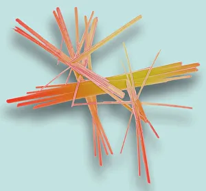
Radiolarian, SEMRadiolarian. Coloured scanning electron micrograph (SEM) of a radiolarian. Radiolarians are amoeboid protozoa that produce intricate mineral skeletons
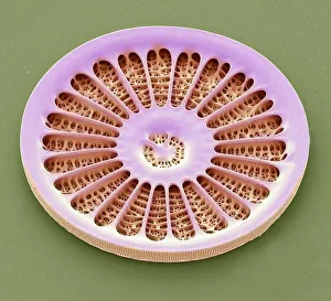
Diatom, SEMDiatom. Coloured scanning electron micrograph (SEM) of a single diatom (Arachnoidiscus sp.). Diatoms may be extremely abundant in both freshwater
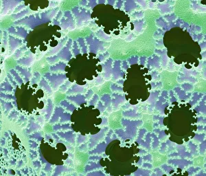
Diatom detail, SEMDiatom. Coloured scanning electron micrograph (SEM) of detail of the silica wall of a diatom. Diatoms may be extremely abundant in both freshwater
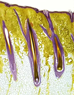
Human skin showing hair follicles, LMHairy skin. Light micrograph of a thick section of human skin, showing three central hair follicles. The outer layer of the skin, the epidermis, is the thin, purple band, supported by the deeper
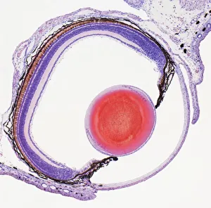
Frog eye, light micrographFrog eye. Light micrograph of a section through the eye of a frog. Most amphibians, such as frogs and salamanders, have color vision
Foraminiferan microfossil, SEMForaminiferan. Coloured scanning electron micrograph (SEM) of a foraminiferan microfossil from maldives beach sand. Microfossils are roughly 0.05 to 2mm in size
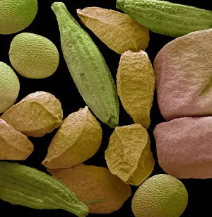
Panch phoran, SEMPanch phoran. Coloured scanning electron micrograph (SEM) of panch phoran or indian five spice. It is a spice blend commonly used in Eastern India and Bangladesh and consists of the following seeds
Sea salt, SEMSea salt. Coloured scanning electron micrograph (SEM) of sea salt, showing its crystalline structure. Sea salt consists mainly of sodium chloride (NaCl), but unlike pure table salt
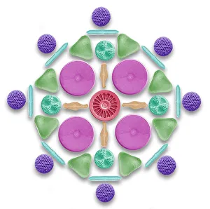
Diatoms and radiolaria, SEMDiatoms and radiolaria. Coloured scanning electron micrograph (SEM) of a circular arrangement of various diatoms and radiolaria. Diatoms are planktonic unicellular algae
Virus particles and bacteria, SEMVaccinia virus particles and bacteria. Coloured scanning electron micrograph (SEM) of vaccinia virus particles (green). Unlike most viruses, vaccinia replicates in the cells cytoplasm
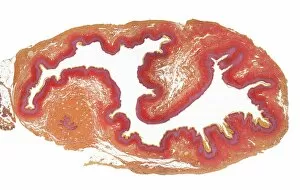
Cervix, LMCervix. Low power lght micrograph (LM) of the cervix. The cervix is the narrow inferior portion of the uterus. The part which projects into the vagina is seen here
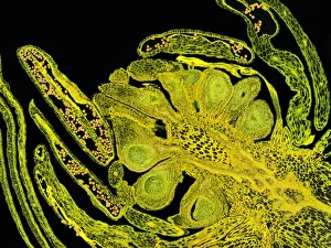
Flower bud, LMFlower bud. Light microscope image (LM) of a section of a flower bud. The small yellow pollen grains are visible in the anthers, the male part of the flower
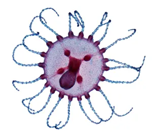
Obelia medusa, LMObelia hydrozoan medusa. Light micrograph (LM) of a medusa (young polyp) from the Obelia geniculata hydroid. The circular shallow semi-bell has solid tentacles on the rim
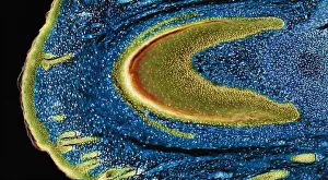
Developing nail, LMDeveloping nail. Light micrograph (LM) of longitudinal section through a fetal finger tip to show the developing nail. The large area of green-yellow nail bed epithelium is tipped by the developing
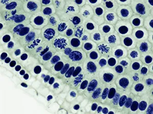
Mitosis, LMMitosis. Light micrograph of onion (Allium cepa) root tip cells undergoing mitosis (nuclear division). Magnification: x600 when printed at 10 centimetres wide
Onion root tip, LMMitosis. Light micrograph (LM) of a transverse section of onion (Allium cepa) root tip to show cells undergoing mitosis (nuclear division). Magnification: x100 when printed at 10 centimetres wide
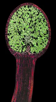
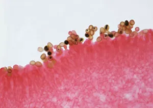
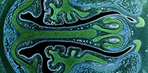
Liverwort spore capsule, LMLiverwort spore capsule. Light micrograph (LM). Longitudinal section through the thallus and sporangium of a liverwort (Pellia epiphylla)
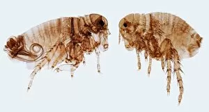
Human fleas, LMHuman fleas. Light micrograph (LM) of a male (left) and female human flea (Pulex irratans). Fleas are wingless and flattened from side to side, which makes them difficult to dislodge in hair
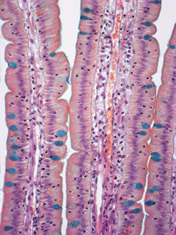
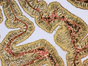
Small intestine, LMSmall intestine. Light micrograph (LM) of a section through the finger-like projections (villi) of the duodenum, the uppermost part of the small intestine
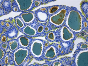
Thyroid, LMThyroid gland. Light micrograph (LM) of a thyroid gland showing the follicles. The follicles are lined by a single layer of cuboidal epithelial cells (blue)
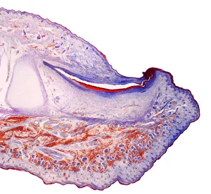
Fingertip, LMFingertip. Light micrograph (LM) of a section through the fingertip. The nail (orange) is at top center, with the nail root below. The nail bed is dark purple and is continous with the epithelium
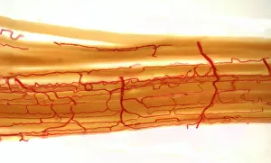
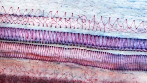
Blood supply to muscles, LMBlood supply to muscles. Light micrograph (LM) showing blood supply to muscle fibers. The muscle fibers (yellow) have been teased apart to reveal the capillary bed (red)
Fallopia tube, LMFallopian tube. Light micrograph (LM).The fallopian tube, or oviduct, conveys the egg from the ovary to the uterus. Ciliated columnar epithelium is yellow
Mushroom gill, LMMushroom gills. High power light micrograph (LM) of a section through the gills of a mushroom, Agaricus sp. (formerly Psalliota sp.)
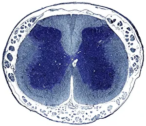
Spinal cord, LMSpinal cord. Light micrograph (LM) of a cross-section through the human spinal cord in the lumbar region. The spinal cord consists of a butterfly-shaped core (dark blue) known as grey matter
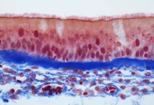
Trachel epithelium, LMTrachea epithelium. Light micrograph (LM) of a vertical section through the pseudostratified columnar epithelium from the trachea
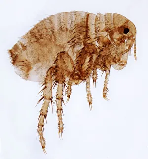
Female flea, LMHuman flea. Light micrograph (LM) of a female human flea (Pulex irratans). Fleas are wingless and flattened from side to side, which makes them difficult to dislodge in hair
Nasal sinuses, LMNasal sinuses. Light micrograph (LM) of the nasal sinuses ( lined by cyan epithelium ) and the supporting cartilages (green). Bone tissue is identified by the blue bone marrow
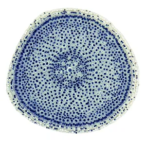
Xylem, LMXylem tissue. Light micrograph (LM) of a section through sunflower(helianthus annuus) tissue showing spiral tracheids, a type of xylem
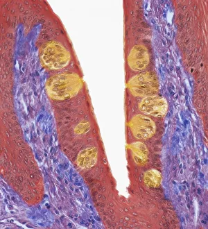
Taste buds, LMTaste buds. Coloured light micrograph of a section through the tongue, showing taste buds (round, yellow). The taste buds are within papillae (projections) located on the surface of the tongue
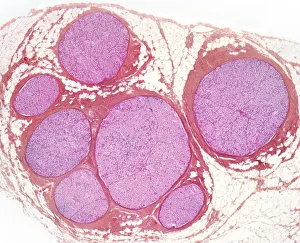
Nerve fibres, LMNerve fibres. Light micrograph (LM) of a transverse section through a bundle (fascicle) of nerve fibres. Within each fascicle are many myelinated nerve fibres
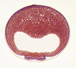
Developing frog egg, LMDeveloping frog egg. Light micrograph of a transverse section through a developing egg laid by a common frog (Rana temporaria). The egg has reached the early neurula stage
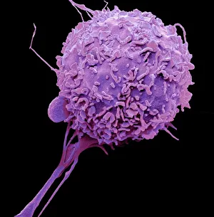
Macrophage, SEMMacrophage. Coloured scanning electron micrograph (SEM) of a macrophage white blood cell. Macrophages are cells of the bodys immune system
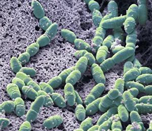
Streptococcus mutans, SEMStreptococcus mutans. Coloured scanning electron micrograph (SEM). S. mutans is a coccoid shaped, Gram-positive, anaerobic bacteria that is part of the normal bacteria flora of the mouth
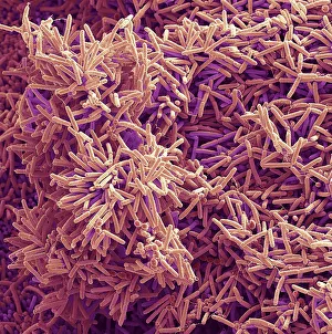
Plaque-forming bacteria, SEMPlaque-forming bacteria, coloured scanning electron micrograph (SEM). Plaque consists of a film of bacteria embedded in a glycoprotein matrix
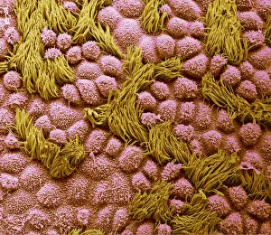
Fallopian tube, SEMFallopian tube. Coloured scanning electron micrograph (SEM) of the surface of a human fallopian tube. Fallopian tubes are ducts that lead from the ovaries to the uterus