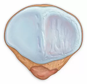
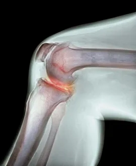
Posterior view patellar surface showing and injured cartilage surface
Horizontal, Detail, Anatomy, Diagram, Illustration, Bone, Injury, Medical, Damaged, Biology, Rear View, Cut Out, White Background, Artwork, Color Image, Close Up, Healthcare And Medicine
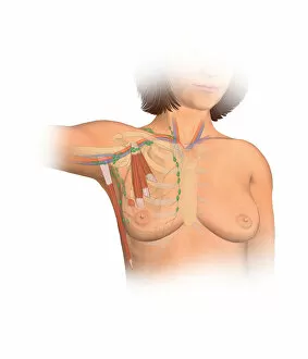
Anterior view female anatomy showing breast tissue with a tumor
Female, Anatomy, Diagram, Muscle, Illustration, Ribs, Bone, Disease, Medical, Vein, Biology, See Through, Cut Out, White Background, Vertical, Artwork, Female Likeness, Breast, Color Image
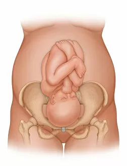
Front view of a woman nine months pregnant (baby phantomed within) ready for delivery
Detail, Baby, Anatomy, Diagram, Mother, Illustration, Bone, Birth, Medical, Fetus, Pregnant, Pregnancy, Biology, Pelvis, Femur, Development, Abdomen, Anterior, See Through, Cut Out, White Background
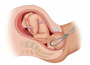
Cross section of the mothers anatomy showing the baby in uteruo LOA being delivered
Horizontal, Detail, Baby, Anatomy, Diagram, Mother, Illustration, Birth, Medical, Fetus, Pregnant, Pregnancy, Uterus, Biology, Development, Abdomen, See Through, Cut Out, White Background, New Life
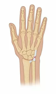
Hand bones with injury
Artwork, Bone, Hand, Hand bones, Human Anatomy, Illustration, Injury, Medical, White Background, Wrist, Anatomy, Vertical, Color Image, No People, biomedical illustration, Healthcare And Medicine
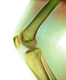
Illustration of the anterior knee, articular surface
anatomy, body part, bone, cartilage, close-up, color image, femur, fibula, front view, human body part, human bone, human joint, human knee, illustration, joint - body part, knee, patella, no people