
Home > Science Inspired Art > The Human Brain

The Human Brain Collection (page 4)

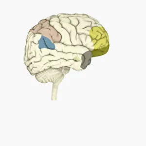
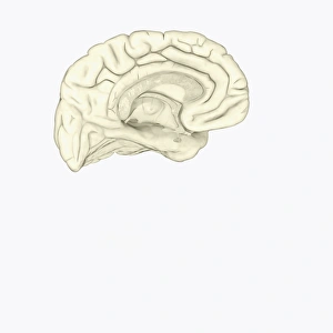
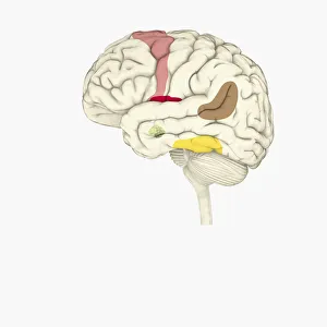
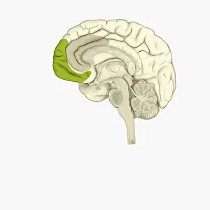
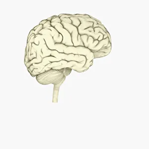
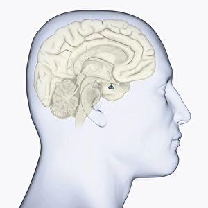
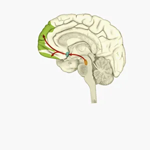
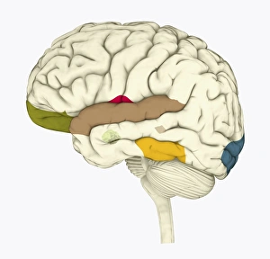
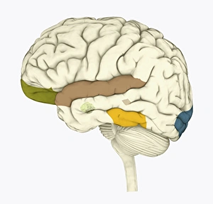
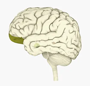
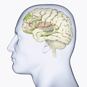
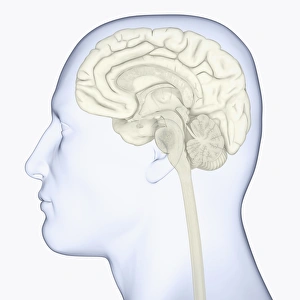
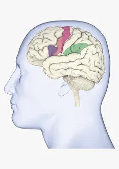
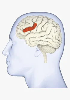
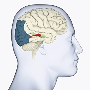
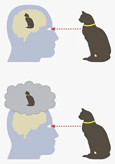
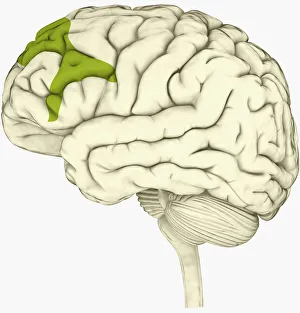
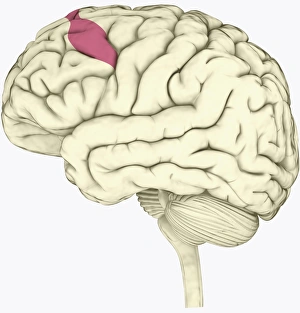
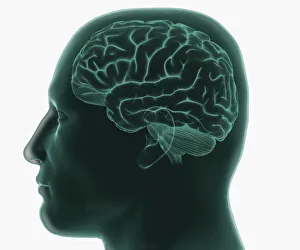
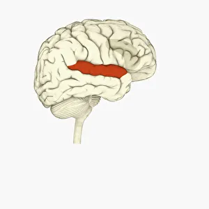
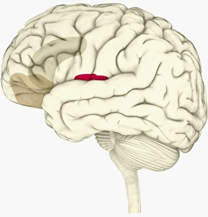
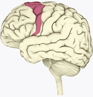
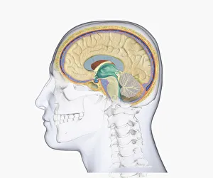
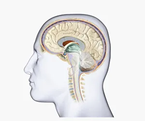
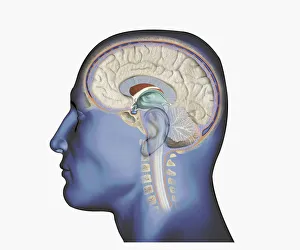
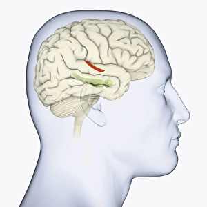
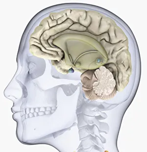
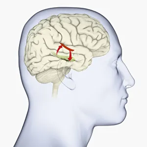
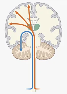
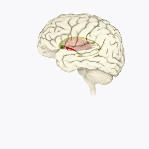
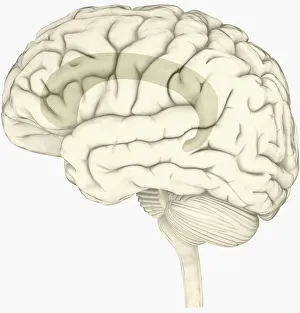
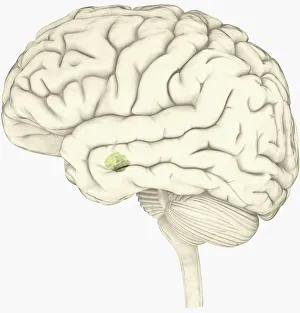
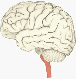
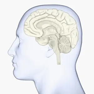
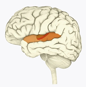
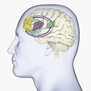
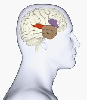
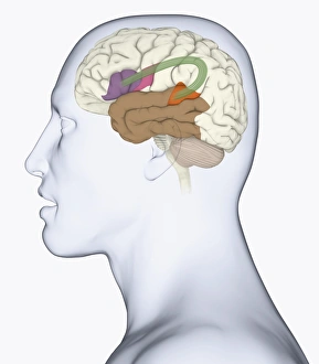
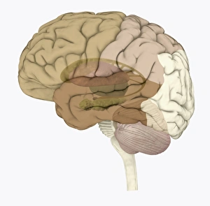
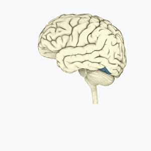
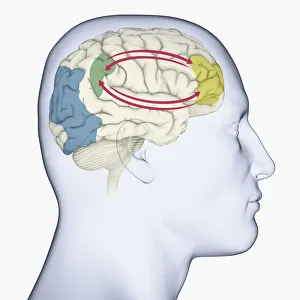
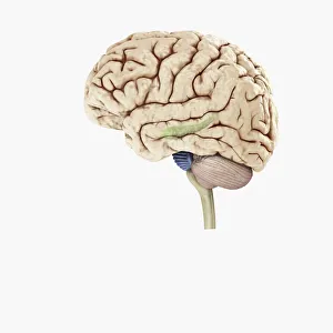
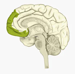
Illustrations of the Human Brain, the command center for the human nervous system and one of the largest and most complex organs in the human body. It is made up of more than 100 billion nerves that communicate in trillions of connections called synapses

213 items

Background imageThe Human Brain Collection: Digital illustration of parietal lobe (green), posterior superior temporal sulcas (blue)

Digital illustration of parietal lobe (green), posterior superior temporal sulcas (blue), temperal pole (grey), dorsolateral prefrontal cortex, amygdala

Background imageThe Human Brain Collection: Digital cross section illustration of human brain

Digital cross section illustration of human brain

Background imageThe Human Brain Collection: Digital illustration of emotional response areas in human brain

Digital illustration of emotional response areas in human brain

Background imageThe Human Brain Collection: Digital illustration of anterior cingulate cortex (grey), and medial frontal cortex (green)

Digital illustration of anterior cingulate cortex (grey), and medial frontal cortex (green) in human brain

Background imageThe Human Brain Collection: Digital illustration of human brain

Digital illustration of human brain

Background imageThe Human Brain Collection: Digital illustration of head in profile showing pituitary gland in brain highlighted in blue

Digital illustration of head in profile showing pituitary gland in brain highlighted in blue

Background imageThe Human Brain Collection: Digital illustration of reward pathway in human brain

Digital illustration of reward pathway in human brain

Background imageThe Human Brain Collection: Digital illustration of human brain associated with full awareness

Digital illustration of human brain associated with full awareness

Background imageThe Human Brain Collection: Digital illustration of areas of information highlighted in human brain

Digital illustration of areas of information highlighted in human brain

Background imageThe Human Brain Collection: Digital illustration of human brain with orbitofrontal cortex and amygdala highlighted in green

Digital illustration of human brain with orbitofrontal cortex and amygdala highlighted in green

Background imageThe Human Brain Collection: Digital illustration of head in profile showing areas of brain used for processing emotion

Digital illustration of head in profile showing areas of brain used for processing emotion

Background imageThe Human Brain Collection: Digital illustration of head in profile showing brain

Digital illustration of head in profile showing brain

Background imageThe Human Brain Collection: Digital illustration of head in profile showing mirror neurons in human brain highlighted in

Digital illustration of head in profile showing mirror neurons in human brain highlighted in purple, pink and green

Background imageThe Human Brain Collection: Digital illustration of head in profile showing area of mirror neuron in brain highlighted in orange

Digital illustration of head in profile showing area of mirror neuron in brain highlighted in orange

Background imageThe Human Brain Collection: Digital illustration of head in profile showing direction of sensory signals from visual cortex to

Digital illustration of head in profile showing direction of sensory signals from visual cortex to hippocampus in brain

Background imageThe Human Brain Collection: Digital illustration of monism and dualism in human brain

Digital illustration of monism and dualism in human brain

Background imageThe Human Brain Collection: Digital illustration of prefrontal cortex of human brain highlighted in green

Digital illustration of prefrontal cortex of human brain highlighted in green

Background imageThe Human Brain Collection: Digital illustration of various areas of cortex in human brain receiving input from sense organs

Digital illustration of various areas of cortex in human brain receiving input from sense organs

Background imageThe Human Brain Collection: Digital illustration of crucial parts of highlighted in human brain

Digital illustration of crucial parts of highlighted in human brain

Background imageThe Human Brain Collection: Digital illustration of frontal lobe and parietal lobe areas (orange) in left hemispheres (blue)

Digital illustration of frontal lobe and parietal lobe areas (orange) in left hemispheres (blue), and pathway of data from parietal lobe to frontal lobe (green) in human brain

Background imageThe Human Brain Collection: Digital illustration of lateral prefrontal cortex involved in decision making highlighted in green

Digital illustration of lateral prefrontal cortex involved in decision making highlighted in green in human brain

Background imageThe Human Brain Collection: Digital illustration of premotor cortex involved in decision making highlighted in pink in human

Digital illustration of premotor cortex involved in decision making highlighted in pink in human brain

Background imageThe Human Brain Collection: Digital illustration of human head in profile showing brain

Digital illustration of human head in profile showing brain

Background imageThe Human Brain Collection: Digital illustration of right superior temporal sulcas, and anterior cingulate cortex highlighted

Digital illustration of right superior temporal sulcas, and anterior cingulate cortex highlighted in red and grey in human brain

Background imageThe Human Brain Collection: Digital illustration of anterior insular, anterior cingulate cortex

Digital illustration of anterior insular, anterior cingulate cortex, and ventromedial prefrontal cortex highlighted in human brain

Background imageThe Human Brain Collection: Digital illustration of premotor cortex involved in decision making highlighted in pink in human

Digital illustration of premotor cortex involved in decision making highlighted in pink in human brain

Background imageThe Human Brain Collection: Digital illustration of head in profile showing skull, brain, and spine

Digital illustration of head in profile showing skull, brain, and spine

Background imageThe Human Brain Collection: Digital illustration of head in profile showing brain and spine

Digital illustration of head in profile showing brain and spine

Background imageThe Human Brain Collection: Digital illustration of head in profile showing brain and spine

Digital illustration of head in profile showing brain and spine

Background imageThe Human Brain Collection: Digital illustration of head in profile showing hippocampus and amygdala (green)

Digital illustration of head in profile showing hippocampus and amygdala (green), and auditory area (red) in brain

Background imageThe Human Brain Collection: Digital illustration of head in profile showing skull, brain and spine

Digital illustration of head in profile showing skull, brain and spine

Background imageThe Human Brain Collection: Digital illustration of head in profile showing auditory cortex (red), and hippocampus (green)

Digital illustration of head in profile showing auditory cortex (red), and hippocampus (green) and direction of signals in brain

Background imageThe Human Brain Collection: Digital illustration of unconscious and conscious pathways in human brain passing through thalamus

Digital illustration of unconscious and conscious pathways in human brain passing through thalamus ending in parietal lobe of cortex

Background imageThe Human Brain Collection: Digital illustration of direction of dopamine flow, nucleus accumbens, basal ganglia

Digital illustration of direction of dopamine flow, nucleus accumbens, basal ganglia and ventral tegmental area highlighted in human brain

Background imageThe Human Brain Collection: Black and white digital illustration of cingulate cortex area of human brain

Black and white digital illustration of cingulate cortex area of human brain

Background imageThe Human Brain Collection: Digital illustration of amygdala in human brain

Digital illustration of amygdala in human brain

Background imageThe Human Brain Collection: Digital illustration of human brain with spine highlighted in pink

Digital illustration of human brain with spine highlighted in pink

Background imageThe Human Brain Collection: Digital illustration of head in profile showing brain

Digital illustration of head in profile showing brain

Background imageThe Human Brain Collection: Digital illustration of human brain with primary auditory cortex highlighted in orange and red

Digital illustration of human brain with primary auditory cortex highlighted in orange and red

Background imageThe Human Brain Collection: Digital illustration of head in profile showing memory areas of brain

Digital illustration of head in profile showing memory areas of brain

Background imageThe Human Brain Collection: Digital illustration of head in profile showing amygdala, auditory cortex, wernickes area

Digital illustration of head in profile showing amygdala, auditory cortex, wernickes area, and anterior temporal lobe in human brain

Background imageThe Human Brain Collection: Digital illustration of head in profile showing bundle of nerve fibres connecti ng Brocas area

Digital illustration of head in profile showing bundle of nerve fibres connecti ng Brocas area and Wernickes area in human brain

Background imageThe Human Brain Collection: Digital illustration of areas associated with memory in human brain

Digital illustration of areas associated with memory in human brain

Background imageThe Human Brain Collection: Digital illustration of human brain

Digital illustration of human brain

Background imageThe Human Brain Collection: Digital illustration of head in profile showing brain and right side of working memory

Digital illustration of head in profile showing brain and right side of working memory

Background imageThe Human Brain Collection: Digital illustration of hippocampus (green) and pons (blue) in left hemisphere of human brain

Digital illustration of hippocampus (green) and pons (blue) in left hemisphere of human brain

Background imageThe Human Brain Collection: Digital illustration of human brain showing frontal cortex in green

Digital illustration of human brain showing frontal cortex in green

Background imageThe Human Brain Collection: Digital illustration of human brain in yellow circle on white background

Digital illustration of human brain in yellow circle on white background


All Professionally Made to Order for Quick Shipping