mail_outline sales@mediastorehouse.com
128 items
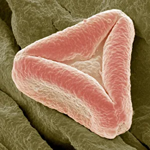
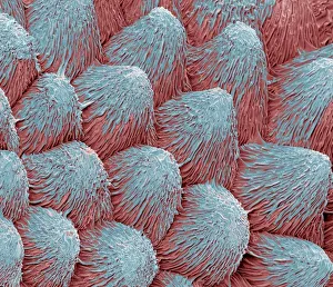
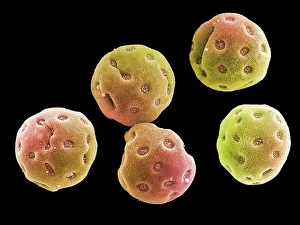
Bottlebrush pollen, SEMBottlebrush (Callistemon) pollen grain, magnified x3000 when printed at 10 centimetres wide
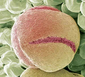
Strawberry pollen, SEMStrawberry (Fragaria vesca) pollen grain, magnified x2000 when printed at 10 centimetres wide
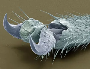
Stick insect foot, SEMClaw of a Stick Insect (Phasmatodea), magnified x80 when printed at 10 centimetres wide
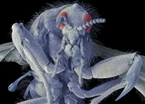
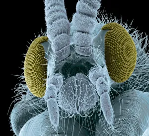
March fly, SEMMarch fly. Scanning electron micrograph (SEM) of a female march fly (family Bibionidae). A member of a family of stout insects in the fly order, Diptera
Orchid petal, SEMOrchid (Phalaenopsis) petal, magnified x450 when printed at 10 centimetres wide
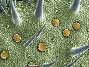
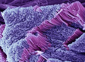
Marjoram leaf surface, scanning electron microscope (SEM)Backgrounds, Biology, Botany, Color Image, Full Frame, Glandular, High, SEM, 85758300
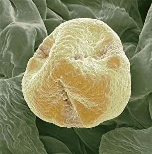
Geranium pollen, SEMGeranium pollen grain, magnified x650 when printed at 10 centimetres wide
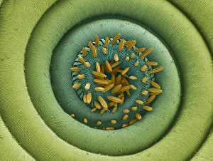
Angels trumpets pollen, SEMAngels Trumpets (Brugmansias) pollen grain, magnified x1600 when printed at 10 centimetres wide
Kiwi fruit pollen grain, SEMkiwi fruit (Actinidia deliciosa) pollen grain, coloured scanning electron micrograph (SEM). Magnification: x3000 when printed at 10 centimetres wide
Scanning electron microscope (SEM) of tendonAnatomy, Biology, Close-Up, Color Image, Connective Tissue, Fiber, Ful, SEM, 85757662
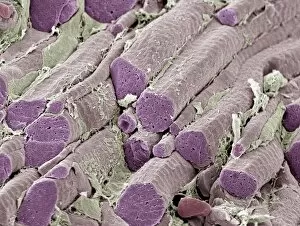
Skeletal muscle fibers, colored scanning electron micrograph (SEM)Anatomy, Backgrounds, Biology, Cartilage, Color Image, Connective Tis, SEM, 85757573
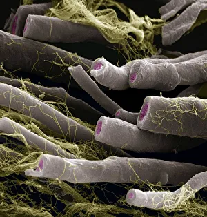
Caterpillar hairs, SEMCaterpillar hairs. Coloured scanning electron micrograph (SEM) of hairs from the vapourer moth (Orgyia antiqua) caterpillar. Magnification: x250 when printed at 10 centimetres tall
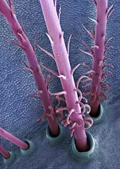
Moth proboscis. Coloured scanning electron micrograph (SEM) of the coiled proboscis of a moth (order Lepidoptera). The proboscis is an elongated part of the mouth
Nerve fibers, scanning electron micrographNerve fibres. Coloured scanning electron micrograph (SEM) of fractured myelinated nerve fibres. The myelin sheath is blue-green
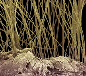
Dog hair, colored scanning electron micrograph (SEM)Dog hair, coloured scanning electron micrograph (SEM). The outside of the hair, the cuticle, is covered in overlapping scales of dead cells containing the protein keratin
Myelinated nerve fibersAxon, Backgrounds, Biology, Color Image, Connective Tissue, Endoneuri, SEM, 85758245
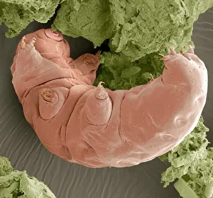
Water bear, SEMWater Bear (Tardigrade), a tiny aquatic invertebrate, magnified x250 when printed at 10 centimetres wide
Osteocyte bone cell, SEMOsteocyte bone cell. Coloured scanning electron micrograph (SEM) of an osteocyte bone cell (blue) surrounded by bone tissue (grey)
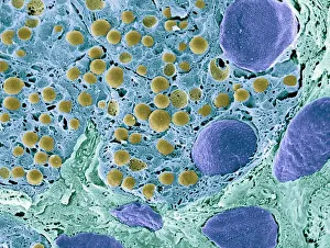
Pancreas tissue, colored scanning electron micrographPancreas tissue. Coloured scanning electron micrograph (SEM) of fractured pancreas tissue. Seen here are zymogen granules (yellow) and cell nuclei (purple)
Bone tissue, close-upAnatomy, Biomedical Illustration, Blue, Close-Up, Color Image, Cross-, SEM, 85757735
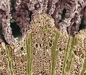
Wood. Scanning electron microscope (SEM)Backgrounds, Biology, Botany, Color Image, Dicotyledon, Full Frame, Hi, SEM, 85757115
Tongue bacteria. Coloured scanning electron micrograph (SEM) of bacteria on the surface of a human tongue. Large numbers of bacteria can form a visible layer on the surface of the tongue
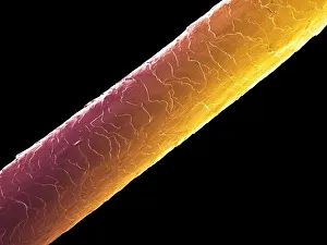
Human hair, semHuman hair (Caucasian, brunette), coloured scanning electron micrograph (SEM). The outer layer of hair (the cuticle) has overlapping scales of keratin
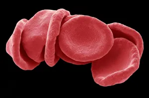
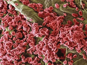
Red blood cells, SEMRed blood cells. Coloured scanning electron micrograph (SEM) of red blood cells (RBCs, erythrocytes). Red blood cells are biconcave
Human blood cells, SEMHuman blood cells. Coloured scanning electron micrograph (SEM) of normal human blood. The majority of the cells are red blood cells, but white blood cells (yellow) and platelets (pink) are also seen
Colored scanning electron micrograph(SEM)Algae, Black Background, Botany, Color Image, Diatom, Frustule, Horizo, SEM, 85758413
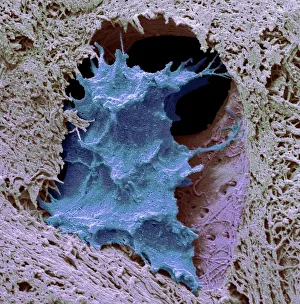
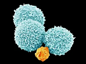
White blood cells, SEMWhite blood cells. Coloured scanning electron micrograph (SEM) of white blood cells (leucocytes). Magnification: x2, 400 when printed at 10 centimetres wide
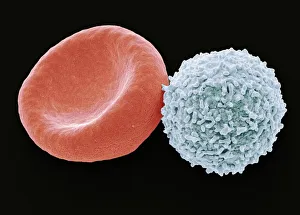
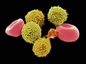
Blood cells, SEMBlood cells. Coloured scanning electron micrograph (SEM) of human blood showing a red and white cell ( lymphocyte). Red blood cells (erythrocytes)
Red blood cells and platelets, SEMRed blood cells and platelets. Coloured scanning electron micrograph (SEM) of human erythrocytes (red blood cells) and a platelet aggregate (orange)
Fly head, colored scanning electron micrographFly head, coloured scanning electron micrograph (SEM). Close-up of the head of a fly, showing its short antennae (upper centre), which are seen between its compound eyes (brown)
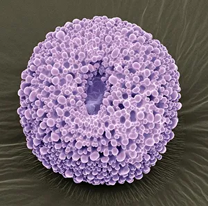
Radiolarian, SEMRadiolarian. Coloured scanning electron micrograph (SEM) of a radiolarian. Radiolarians are amoeboid protozoa that produce intricate mineral skeletons
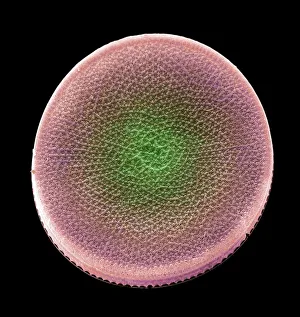
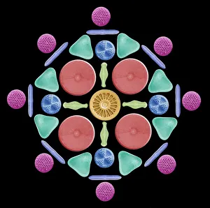
Diatoms and radiolaria, SEMDiatoms and radiolaria. Coloured scanning electron micrograph (SEM) of a circular arrangement of various diatoms and radiolaria. Diatoms are planktonic unicellular algae
Blood supply to muscles, LMBlood supply to muscles. Light micrograph (LM) showing blood supply to muscle fibers. The muscle fibers (blue) have been teased apart to reveal the capillary bed (cyan)
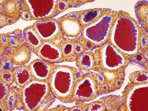
Thyroid, LMThyroid gland. Light micrograph (LM) of a thyroid gland showing the follicles. The follicles are lined by a single layer of cuboidal epithelial cells (orange)
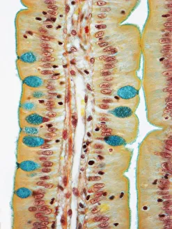
Small intestine, LMSmall intestine. Light micrograph (LM) of a section through the finger-like projections (villi) of the duodenum, the uppermost part of the small intestine
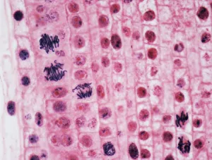
Mitosis, LMMitosis. Light micrograph of onion (Allium cepa) root tip cells undergoing mitosis (nuclear division). Magnification: x600 when printed at 10 centimetres wide
Convolvulus pollen grains, SEMConvolvulus pollen grains. Coloured scanning electron micrograph (SEM) of pollen grains from a convolvulus flower. Convolvulus is a genus of about 200 to 250 species of flowering plants in
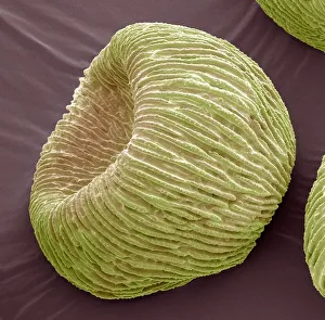
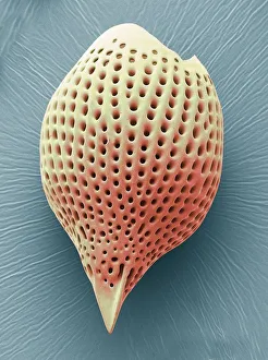
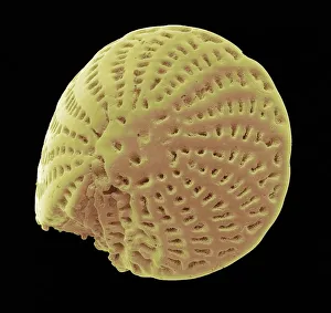
Foraminiferan microfossil, SEMForaminiferan. Coloured scanning electron micrograph (SEM) of a foraminiferan microfossil from maldives beach sand. Microfossils are roughly 0.05 to 2mm in size
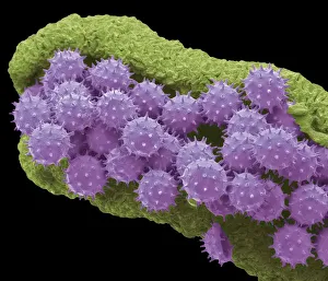
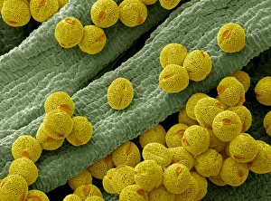
Hibiscus pollen grains, SEMChinese hibiscus pollen. Coloured scanning electron micrograph (SEM) of pollen grains on the anther of a Chinese hibiscus (Hibiscus rosa-sinensis) flower
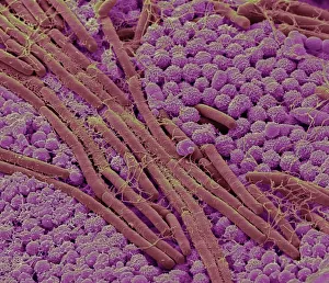
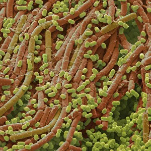
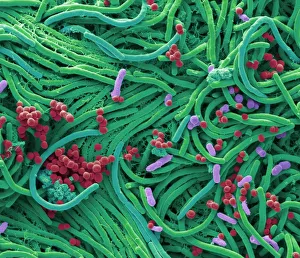
Soil bacteria, SEMSoil bacteria. Coloured scanning electron micrograph (SEM). Bacteria in the soil are directly tied to nutrient recycling especially carbon, nitrogen, phosphorus and sulfur
Verbena pollen, SEMVerbena pollen. Coloured scanning electron micrograph (SEM) of pollen grains from verbena bonariensis. Verbena bonariensis is a tall, slender-stemmed perennial
Tendon, SEMTendon, coloured scanning electron micrograph (SEM), showing bundles of collagen fibres. The parallel alignment of the fibres make tendons inelastic but flexible. Tendons attach muscle to bone
Bellflower pollen, SEMBellflower pollen. Coloured scanning electron micrograph (SEM) of pollen grains from a bellflower (Campanula sp.). Pollen grains are the male gametes (sex cells) of a plant
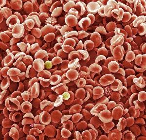
Blood cells, SEMBlood cells. Coloured scanning electron micrograph (SEM) of human red blood cells (erythrocytes, red), white blood cells (leukocytes, yellow), and platelets (thrombocyte, orange)
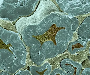
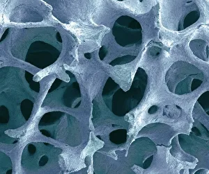
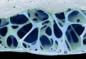
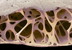
Bird bone tissue, SEMBird bone tissue. Coloured scanning electron micrograph (SEM) of cancellous (spongy) bone from a starlings (Sturnus vulgaris) skull
Red blood cells, SEMRed blood cells, coloured scanning electron micrograph (SEM). Magnification: x1000 when printed at 10 centimetres wide
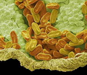
Bacteria found on mobile phone, SEMBacteria found on mobile phone. Coloured scanning electron micrograph (SEM) of bacteria cultured from a mobile phone. Tests have revealed the average handset carries 18 times more potentially harmful