mail_outline sales@mediastorehouse.com
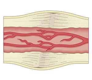
Cross section biomedical illustration of bone repairing itself with dense, compact bone gradually replacing the callus, and blood vessel regrowth
Cross section biomedical illustration of site of incision for microdiscectomy surgical procedure
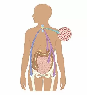
Cross section biomedical illustration of how oral and non-oral drugs circulate in the body, showing liver and inset
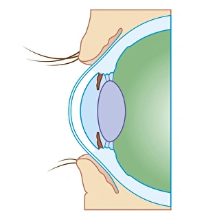
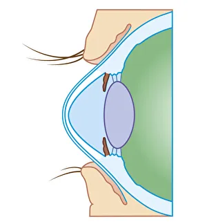
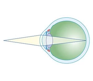
Cross section biomedical illustration of normal eye
Cross section biomedical illustration of keratoconus or conical cornea
Cross section biomedical illustration of Peritoneal Dialysis using the peritoneum as a filter with catheter inserted through abdomen
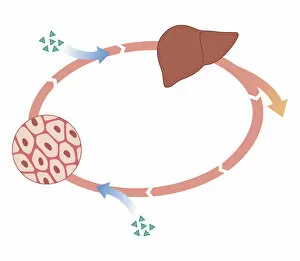
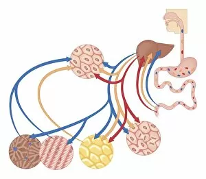
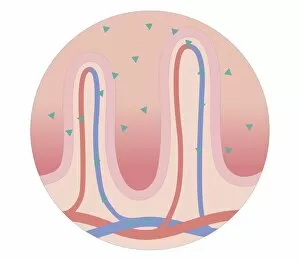
Cross section biomedical illustration of how the body uses food for energy, the liver for digestion, fat storage and glycogen storage
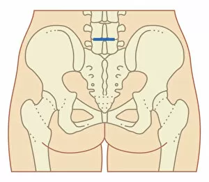
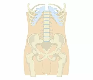
Cross section biomedical illustration of rib cage, spine and pelvis, and femur of adult male
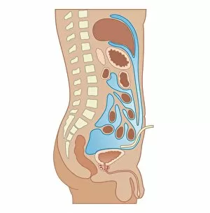
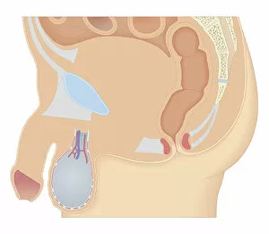
Cross section biomedical illustration of male reproductive system
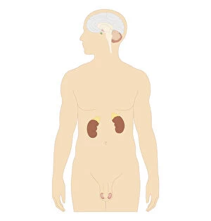
Cross section biomedical illustration of man endocrine system
Cross section biomedical illustration of female anus and rectum
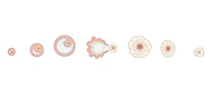
Cross section biomedical illustration of egg development inside ovary during human menstrual cycle
Cross section biomedical illustration of thickening of endometrium during menstrual cycle
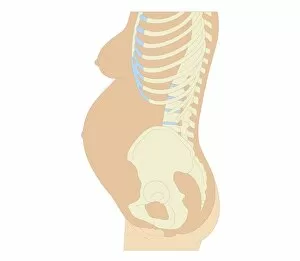
Cross section biomedical illustration of anatomy at 36 weeks pregnant
Cross section biomedical illustration of female reproductive organs six weeks after childbirth
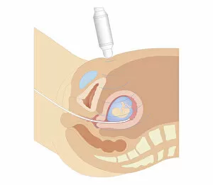
Cross section biomedical illustration of chorionic villus sampling procedure
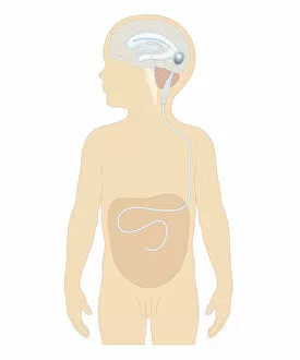
Cross section biomedical illustration of cerebral shunt with valve inserted in brain of boy to remove excess cerebrospinal fluid with tube to carry into stomach
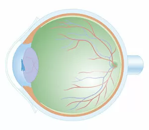
Cross section biomedical illustration of anatomy of human eye
Cross section biomedical illustration of testis in teenage boy
Cross section biomedical illustration of intravenous route of drugs injected into blood vessel for rapidly circulation
Cross section biomedical illustration of oral route of drugs swallowed and passing into digestive system
Cross section biomedical illustration of thyroid and parathyroid hormones in adult female
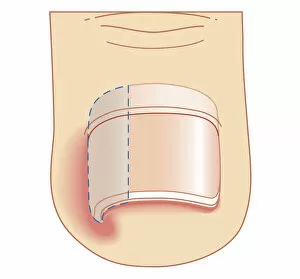
Cross section biomedical illustration of removal site of ingrown toenail (Onychocryptosis)
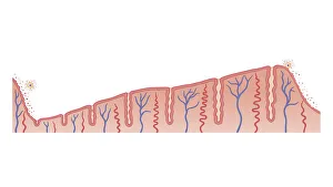
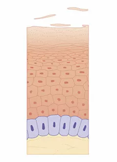
Cross section biomedical illustration of skin growth showing four layers of epidermis
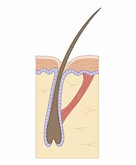
Cross section biomedical illustration of telogen (resting) phase of hair follicle, and arrectores pilorum muscle
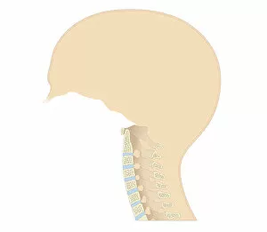
Cross section biomedical illustration of vertebral column in neck showing cervical curve and cervical vertebrae, close-up
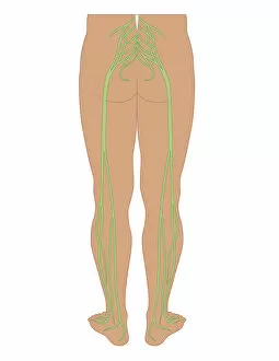
Cross section biomedical illustration of sciatic nerves beginning at the lower back, through buttocks down to lower limbs
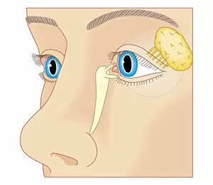
Cross section biomedical illustration of position of lacrimal duct and lacrimal gland
Cross section biomedical illustration of eye focusing on near object
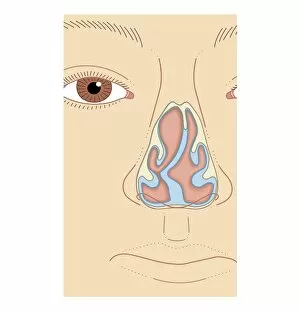
Cross section biomedical illustration of nasal septum deviation, close-up
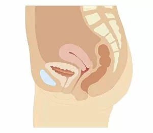
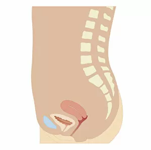
Cross section biomedical illustration of uterus, bladder and pubic bone in adult female
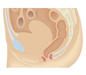
Cross section biomedical illustration of human male reproductive system and pelvis
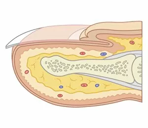
Cross section biomedical illustration of fingernail
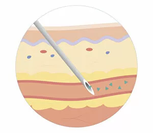
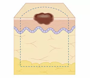
Cross section biomedical illustration of site of skin biopsy
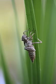
Exuvia, remaining larval skin, cast skin of a dragonfly, old skin after moulting
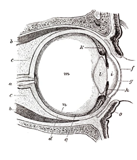
Eye, anatomical illustration
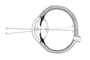
Eye, schematic representation
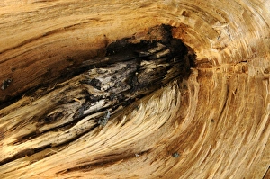
Wood structure
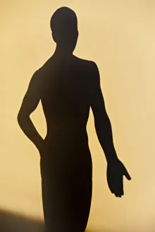
Acupuncture figure as a shadow
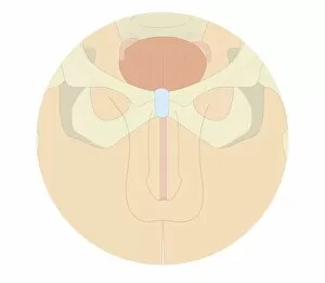
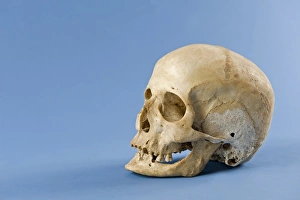
Human skull
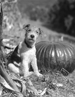
Wire Hair TerrierUNITED STATES - CIRCA 1930s: Portrait Of A Dog, A Wire Hair Terrier Sitting By Pumpkin And Looking At Camera, Making Eye Contact And Looking Cute Outdoors In The Autumn. (Photo by H)
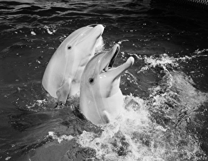
Dolphins jumping out of waterUNITED STATES - CIRCA 1950s: Two dolphins jumping out of water, one with mouth open. (Photo by H. Armstrong Roberts/Retrofile/Getty Images)
Dog holding newspaperUNITED STATES - CIRCA 1950s: Mixed breed dog holding newspaper in mouth. (Photo by H. Armstrong Roberts/Retrofile/Getty Images)