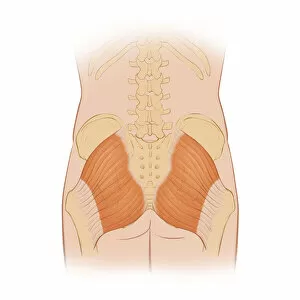
Normal posterior view of the hip bones and gluteus maximus muscle including the lumbar
Detail, Anatomy, Diagram, Muscle, Illustration, Bone, Medical, Hip, Biology, Pelvis, Femur, See Through, Rear View, Cut Out, White Background, Artwork, Color Image, Close Up, Healthcare And Medicine
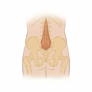
Normal posterior view of the back highlighting the multifidus muscle
Detail, Anatomy, Diagram, Muscle, Illustration, Ribs, Bone, Medical, Biology, Pelvis, See Through, Rear View, Cut Out, White Background, Artwork, Color Image, Close Up, Healthcare And Medicine
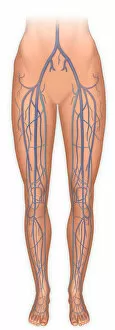
Anterior view of legs and the veinous system
Detail, Anatomy, Diagram, Illustration, Standing, Leg, Barefoot, Medical, Vein, Biology, See Through, Cut Out, White Background, Vertical, Artwork, Low Section, Color Image, Front View, Close Up