mail_outline sales@mediastorehouse.com
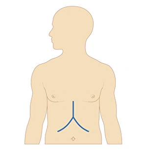
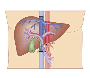
Biomedical illustration of incision site for liver transplant
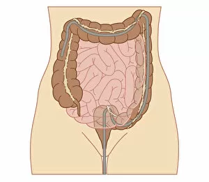
Cross section biomedical illustration of colonoscopy procedure in adult female
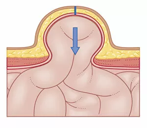
Cross section biomedical illustration of hernia repair and site of incision to uncover the hernia
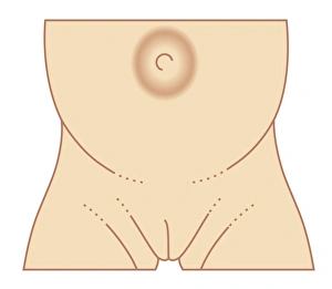
Biomedical illustration of umbilical hernia in female baby
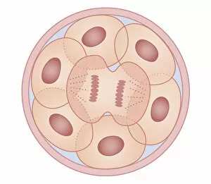
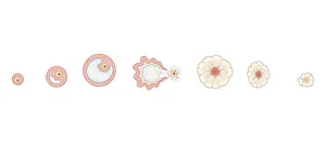
Cross section biomedical illustration of human cell division with zygote dividing to form new cells
Cross section biomedical illustration of cervical cap in position
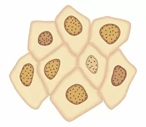
Cross section biomedical illustration of cancer cells forming tumour at third doubling stage
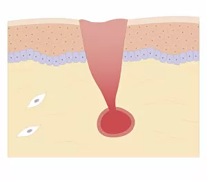
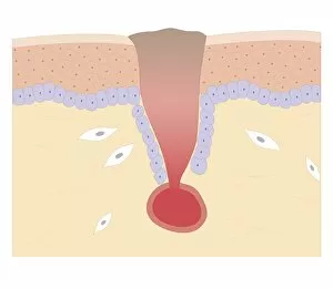
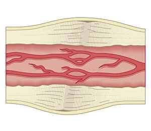
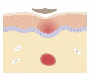
Cross section biomedical illustration of damaged skin and blood vessel
Cross section biomedical illustration of blood clotting over broken skin and fibroblasts multiplying and migrating to injury
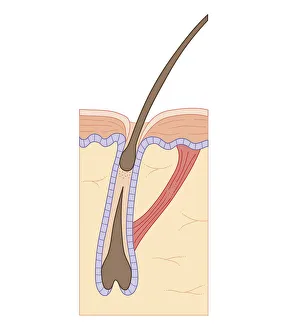
Cross section biomedical illustration of hair follicle during growth phase
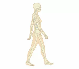
Biomedical illustration of adult male
Cross section biomedical illustration of adult female human skeletal system and joints
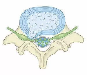
Cross section biomedical illustration of lumber microdisectomy before surgery
Cross section biomedical illustration of liver transplant procedure
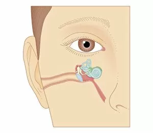
Cross section biomedical illustration of internal components of the ear
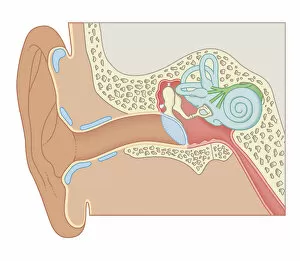
Cross section biomedical illustration of the anatomy of the ear
Cross section biomedical illustration of bone repairing itself with dense, compact bone gradually replacing the callus, and blood vessel regrowth
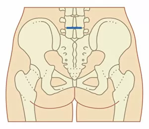
Cross section biomedical illustration of site of incision for microdiscectomy surgical procedure
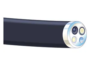
Biomedical illustration of tip of flexible endoscope
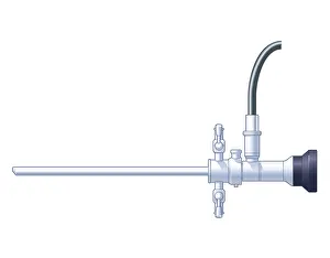
Biomedical illustration of rigid endoscope
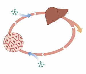
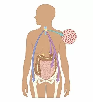
Cross section biomedical illustration of how oral and non-oral drugs circulate in the body, showing liver and inset
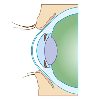
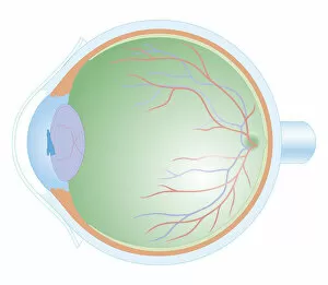
Cross section biomedical illustration of normal eye
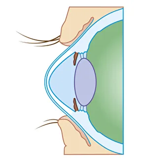
Cross section biomedical illustration of keratoconus or conical cornea
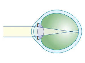
Cross section biomedical illustration of myopia
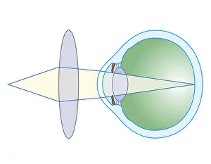
Cross section biomedical illustration of lens to correct hypermetropia
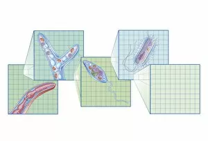
Cross section biomedical illustration on grid of Worm, Fungi, Protozoa, and Bacteria infection and infestation
Cross section biomedical illustration of Peritoneal Dialysis using the peritoneum as a filter with catheter inserted through abdomen
Biomedical illustration of Otoacoustic Emission sound wave chart
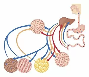
Cross section biomedical illustration of how the body uses food for energy, the liver for digestion, fat storage and glycogen storage
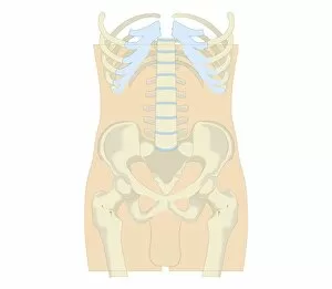
Cross section biomedical illustration of rib cage, spine and pelvis, and femur of adult male
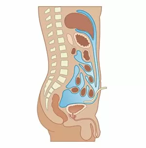
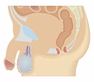
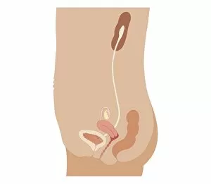
Cross section biomedical illustration of male reproductive system
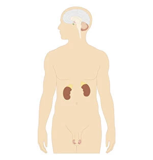
Cross section biomedical illustration of man endocrine system
Cross section biomedical illustration of female anus and rectum
Cross section biomedical illustration of egg development inside ovary during human menstrual cycle
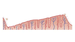
Cross section biomedical illustration of thickening of endometrium during menstrual cycle
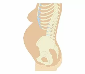
Cross section biomedical illustration of anatomy at 36 weeks pregnant
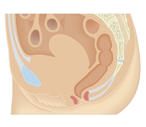
Cross section biomedical illustration of female reproductive organs six weeks after childbirth
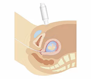
Cross section biomedical illustration of chorionic villus sampling procedure
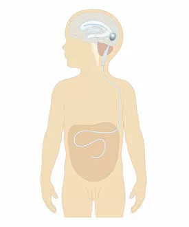
Cross section biomedical illustration of cerebral shunt with valve inserted in brain of boy to remove excess cerebrospinal fluid with tube to carry into stomach
Cross section biomedical illustration of anatomy of human eye
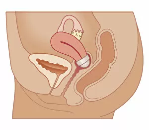
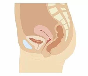
Cross section biomedical illustration of urinary and reproductive systems in adult female
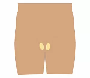
Cross section biomedical illustration of testis in teenage boy
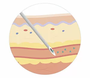
Cross section biomedical illustration of intravenous route of drugs injected into blood vessel for rapidly circulation
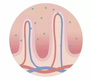
Cross section biomedical illustration of oral route of drugs swallowed and passing into digestive system
Cross section biomedical illustration of thyroid and parathyroid hormones in adult female
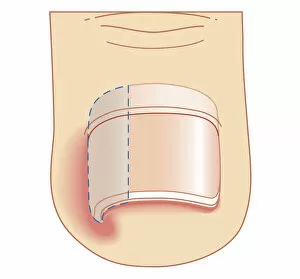
Cross section biomedical illustration of removal site of ingrown toenail (Onychocryptosis)
Cross section biomedical illustration of skin repair with fibrous plug hardening to become scab which falls off when new skin growth is complete
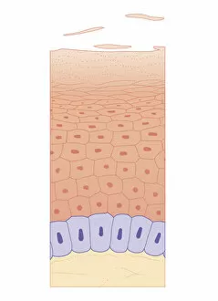
Cross section biomedical illustration of skin growth showing four layers of epidermis