mail_outline sales@mediastorehouse.com
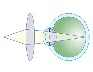
Cross section biomedical illustration of lens to correct hypermetropia
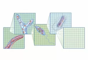
Cross section biomedical illustration on grid of Worm, Fungi, Protozoa, and Bacteria infection and infestation
Cross section biomedical illustration of Peritoneal Dialysis using the peritoneum as a filter with catheter inserted through abdomen
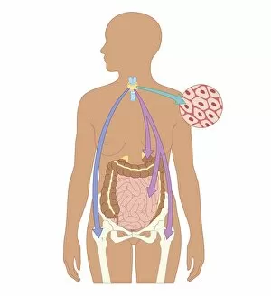
Biomedical illustration of Otoacoustic Emission sound wave chart
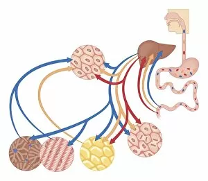
Cross section biomedical illustration of how the body uses food for energy, the liver for digestion, fat storage and glycogen storage
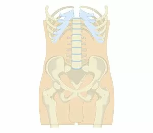
Cross section biomedical illustration of rib cage, spine and pelvis, and femur of adult male
Cross section biomedical illustration of male reproductive system
Cross section biomedical illustration of man endocrine system
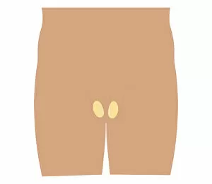
Cross section biomedical illustration of female anus and rectum
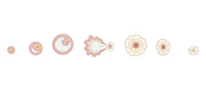
Cross section biomedical illustration of egg development inside ovary during human menstrual cycle
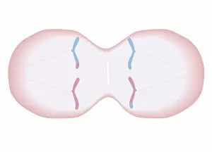
Cross section biomedical illustration of thickening of endometrium during menstrual cycle
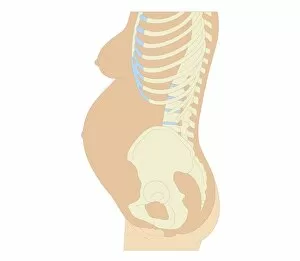
Cross section biomedical illustration of anatomy at 36 weeks pregnant
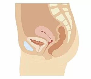
Cross section biomedical illustration of female reproductive organs six weeks after childbirth
Cross section biomedical illustration of chorionic villus sampling procedure
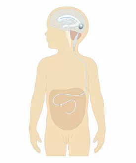
Cross section biomedical illustration of cerebral shunt with valve inserted in brain of boy to remove excess cerebrospinal fluid with tube to carry into stomach
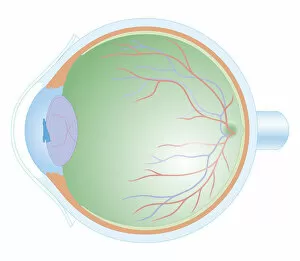
Cross section biomedical illustration of anatomy of human eye
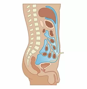
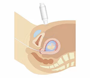
Cross section biomedical illustration of urinary and reproductive systems in adult female
Cross section biomedical illustration of testis in teenage boy
Cross section biomedical illustration of intravenous route of drugs injected into blood vessel for rapidly circulation
Cross section biomedical illustration of oral route of drugs swallowed and passing into digestive system
Cross section biomedical illustration of thyroid and parathyroid hormones in adult female
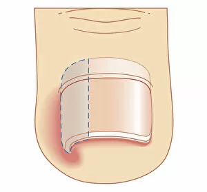
Cross section biomedical illustration of removal site of ingrown toenail (Onychocryptosis)
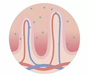
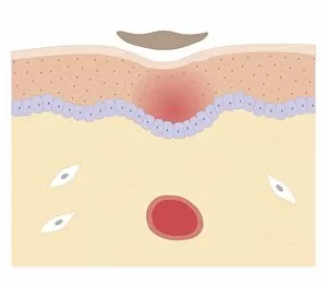
Cross section biomedical illustration of skin repair with fibrous plug hardening to become scab which falls off when new skin growth is complete
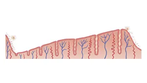
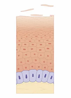
Cross section biomedical illustration of skin growth showing four layers of epidermis
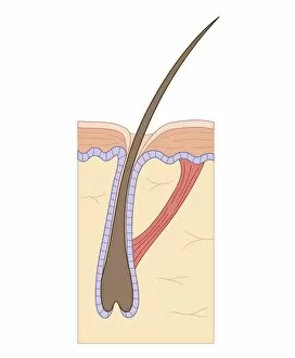
Cross section biomedical illustration of telogen (resting) phase of hair follicle, and arrectores pilorum muscle
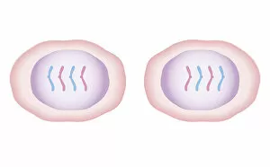
Cross section biomedical illustration of mitosis where nucleus membrane form around each set of chromosomes and the cell begins to divide in two
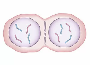
Cross section biomedical illustration of mitosis with new cells form, each having a central nucleus containing identical set of chromosomes
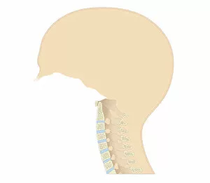
Cross section biomedical illustration of vertebral column in neck showing cervical curve and cervical vertebrae, close-up
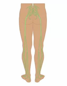
Cross section biomedical illustration of sciatic nerves beginning at the lower back, through buttocks down to lower limbs
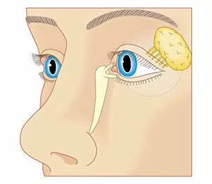
Cross section biomedical illustration of position of lacrimal duct and lacrimal gland
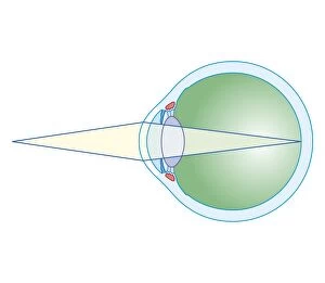
Cross section biomedical illustration of eye focusing on near object
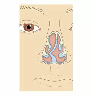
Cross section biomedical illustration of nasal septum deviation, close-up
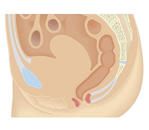
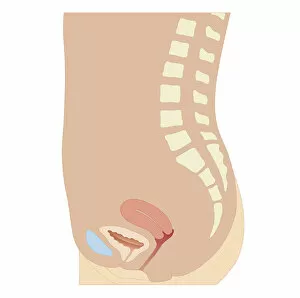
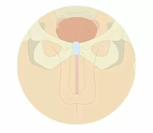
Cross section biomedical illustration of uterus, bladder and pubic bone in adult female
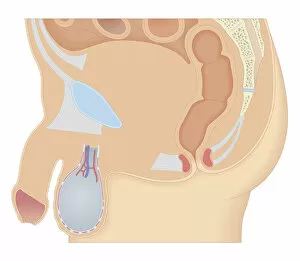
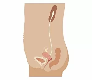
Cross section biomedical illustration of human male reproductive system and pelvis
Cross section biomedical illustration of meiosis with duplicated chromosomes lined up and more threads attach, pulling the duplicated chromosomes apart to form two single chromosomes
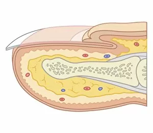
Cross section biomedical illustration of fingernail
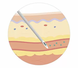
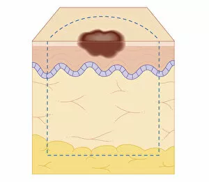
Cross section biomedical illustration of site of skin biopsy
Biomedical illustration of X Chromosome
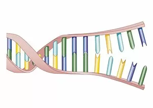
Biomedical illustration of DNA Replication
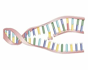
Biomedical illustration of protein synthesis within DNA
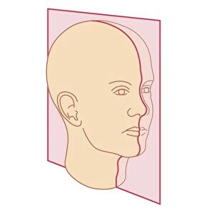
Cross section biomedical illustration of MRI scan plane
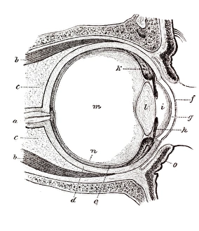
Eye, anatomical illustration
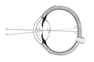
Eye, schematic representation
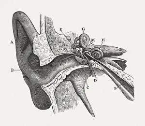
The human ear, wood engraving, published in 1880Anatomy of the human ear: A) auricle, B) External Auditory Canal, C) Tympanic Membrane, D) Tympanic Cavity, E) Malleus, M) Incus, H) Cochlea, G) Semicircular Canals, I) Eustachian Tube
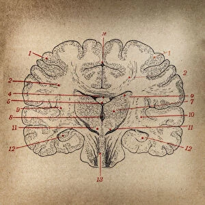
Brain section
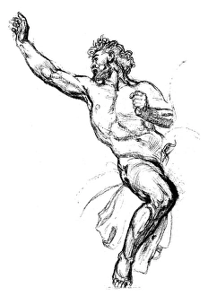
Antique illustration of sketch of Attilas bodyAntique illustration of sketch of Attilas naked body, the character of a Pierre Corneilles drama, 17th century French tragedian