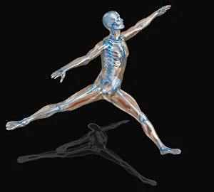
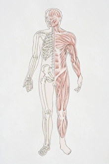
Digitally generated image of human representation dancing ballet
digitally generated image, human representation, dancing, motion, action, freedom, leap, anatomy, internal organ, human skeleton, human bone, free, escape, unleashed, reaching, striving, aspiration
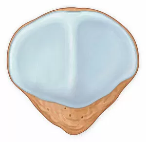
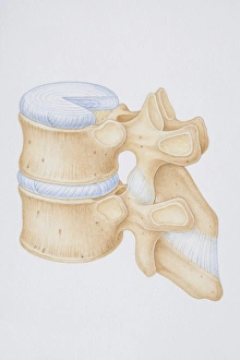
Posterior view patellar surface showing normal cartilage
Horizontal, Detail, Anatomy, Diagram, Illustration, Bone, Medical, Biology, Rear View, Cut Out, White Background, Artwork, Color Image, Close Up, Healthcare And Medicine, The Human Body, No People
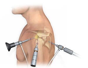
Lateral view of arthroscopic surgical repair on the shoulder joint
Horizontal, Detail, Anatomy, Diagram, Repair, Illustration, Bone, Shoulder, Equipment, Medical, Tool, Biology, Surgery, Joint, See Through, Cut Out, White Background, Artwork, Male Likeness
Open shoulder joint showing inflamed bursa from a bone spur and torn labrum
Detail, Anatomy, Diagram, Illustration, Bone, Shoulder, Injury, Medical, Biology, Joint, Torn, Injured, Cut Out, White Background, Vertical, Artwork, Color Image, Close Up, Healthcare And Medicine
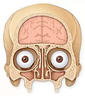
Normal coronal section of the skull and brain showing the coronal sinuses
Brain, Anatomy, Diagram, Illustration, Bone, Skull, Medical, Eyeball, Biology, See Through, Cut Out, White Background, Eye, Vertical, Artwork, Color Image, Front View, Human Skull
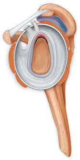
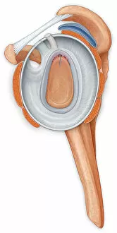
Lateral view of the shoulder joint showing the repair to the labrum
Detail, Anatomy, Diagram, Muscle, Illustration, Bone, Shoulder, Medical, Biology, Joint, Cut Out, White Background, Vertical, Artwork, Color Image, Close Up, Healthcare And Medicine, The Human Body
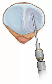
Posterior view patellar surface showing injured cartilage being cleaned up with a shaver
Detail, Anatomy, Diagram, Illustration, Bone, Injury, Medical, Tool, Damaged, Damage, Biology, Surgery, Improvement, Rear View, Cut Out, White Background, Vertical, Artwork, Color Image, Close Up
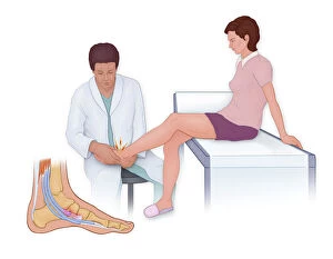
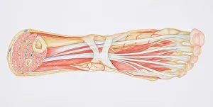
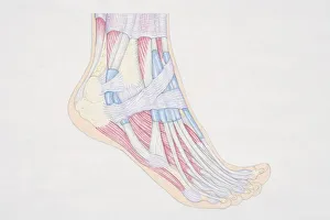
Accessory Navicular Pain Syndrome is caused by friction
Horizontal, Anatomy, Diagram, Illustration, Foot, Bone, Pressure, Injury, Medical, Doctor, Physician, Patient, Biology, Joint, Side View, See Through, Cut Out, White Background, Artwork
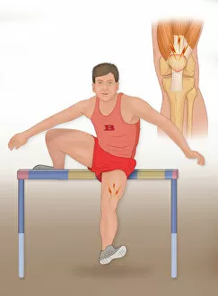
A leg and knee injury sustained from a repetitive motion
Fitness, Sport, Vest, Anatomy, Diagram, Muscle, Illustration, Bone, Repetition, Athlete, Shorts, Leg, Injury, Medical, Jumping, Biology, Knee, See Through, Weekend Activities, Hurdle
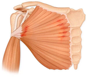
Microscopic muscle fiber tears of the pectoralis major muscle
Artwork, Bone, Clavicle, Human Anatomy, humerus, Illustration, inflammation, Injury, Medical, Microscopic, Muscle, muscle fiber tear, Pectoralis major muscle, Pectoralis Major Tear Syndrome, scapula
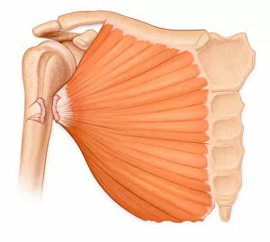
Complete rupture of the pectoralis major tendon from the humerus
Horizontal, Detail, Anatomy, Diagram, Muscle, Illustration, Bone, Injury, Medical, Biology, Bicep, Cut Out, White Background, Artwork, Color Image, Close Up, Healthcare And Medicine, The Human Body
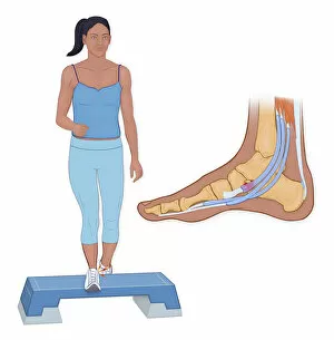
Os Trigonum Pain Syndrome is associated with high impact force or activites on the foot
Fitness, Exercise, Anatomy, Diagram, Pain, Illustration, Bone, Injury, Medical, Biology, Sportswear, Stepping, See Through, Weekend Activities, Cut Out, White Background, Lifestyle, Vertical
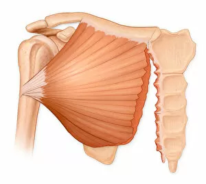
Pectoralis muscle tear, associated with a hematoma formation and cosmetic deformity
Horizontal, Detail, Anatomy, Diagram, Muscle, Illustration, Bone, Injury, Medical, Biology, Cut Out, White Background, Artwork, Color Image, Close Up, Healthcare And Medicine, The Human Body
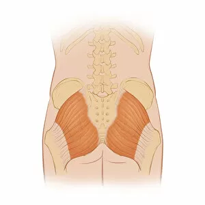
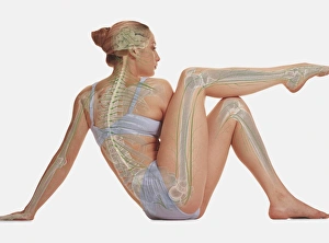
Normal posterior view of the hip bones and gluteus maximus muscle including the lumbar
Detail, Anatomy, Diagram, Muscle, Illustration, Bone, Medical, Hip, Biology, Pelvis, Femur, See Through, Rear View, Cut Out, White Background, Artwork, Color Image, Close Up, Healthcare And Medicine
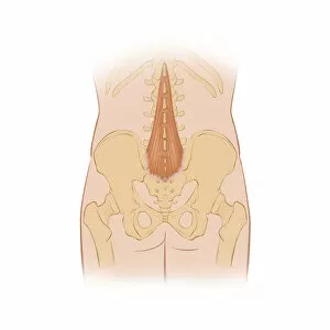
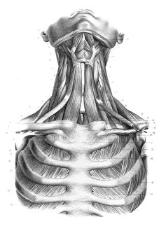
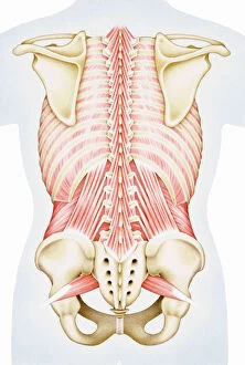
Normal posterior view of the back highlighting the multifidus muscle
Detail, Anatomy, Diagram, Muscle, Illustration, Ribs, Bone, Medical, Biology, Pelvis, See Through, Rear View, Cut Out, White Background, Artwork, Color Image, Close Up, Healthcare And Medicine
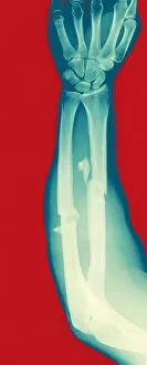
Anatomy, Biology, Broken, Close-Up, Color Image, Colored Background, Forearm, Fracture, Healthcare And Medicine, Human Arm, Human Bone, Human Figure, Human Hand, Photography, Radius, The Human Body