mail_outline sales@mediastorehouse.com
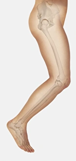
Diagram showing bones inside human leg, leaping forward
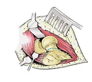
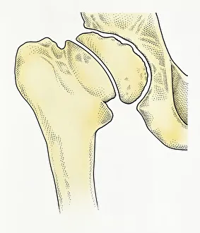
Diagram showing removal of affected head of a femur bone
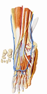
Human foot, internal anatomy
Illustration showing formation of human bone
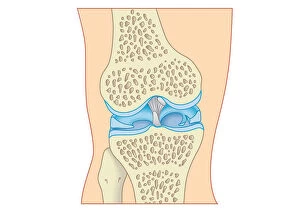
Digital cross section illustration of torn cartilage in knee
Digital illustration showing degeneration of hip joint known as Perthes disease
Illustration showing Osteoporosis in human bone
Cross section illustration of human finger
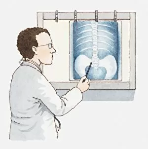
Illustration of doctor pointing at x-ray of pelvis and spine
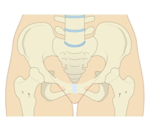
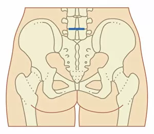
Cross section biomedical illustration of female type pelvis
Cross section biomedical illustration of epidural anaesthesia during labour
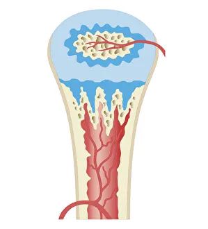
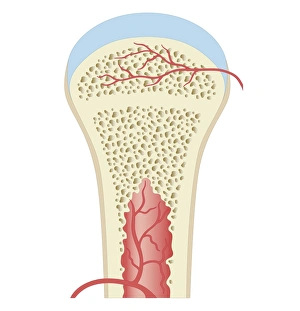
Cross section biomedical illustration of long bone of newborn baby
Cross section biomedical illustration of childhood development long bone
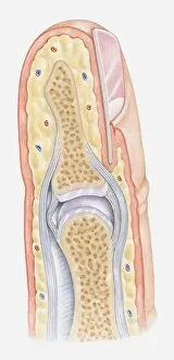
Cross section biomedical illustration of fully developed long bone in adult
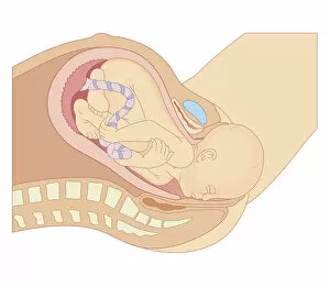
Cross section biomedical illustration of birth of baby with head emerging from vagina
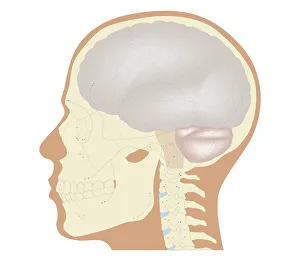
Cross section biomedical illustration of the brain and skull at 18 years of age
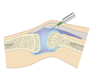
Cross section biomedical illustration of removing synovial fluid from knee using joint aspiration
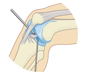
Cross section biomedical illustration of inside the knee joint during rigid endoscopy procedure
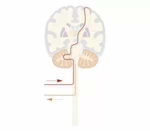
Cross section biomedical illustration of voluntary pathway of human brain
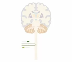
Cross section biomedical illustration of spinal reflex pathway of human brain
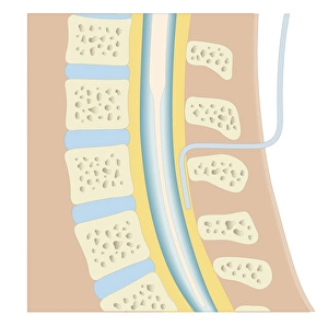
Cross section biomedical illustration of lumber microdisectomy before surgery
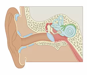
Cross section biomedical illustration of the anatomy of the ear
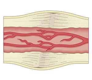
Cross section biomedical illustration of bone repairing itself with dense, compact bone gradually replacing the callus, and blood vessel regrowth
Cross section biomedical illustration of site of incision for microdiscectomy surgical procedure
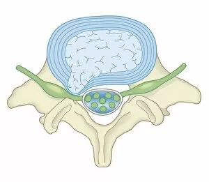
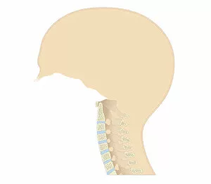
Cross section biomedical illustration of vertebral column in neck showing cervical curve and cervical vertebrae, close-up
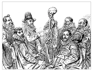
Antique illustration of 17th century anatomy lesson with skeleton: Doctor Egberts teach his students the skeleton structure (from a painting by the 17th century Dutch painter Thomas de Keyser)