mail_outline sales@mediastorehouse.com
Choose a picture from our Images Dated 8th January 2019 Collection for your Wall Art and Photo Gifts
15 items
Richard and Cosima Wagner with Liszt and Hans von Wolzogen in their home WahnfriedIllustration from 19th century
Holland, Haarlem - Iconic WindmillView of the Windmill De Adriaan - one of the iconic landmarks of the city of Haarlem in the Netherlands
Double rainbow landscape in beautiful Irish landscape scenery. Co Tipperary IrelandDouble rainbow landscape in beautiful Irish landscape scenery, taken on sunny and rainy day.Co Tipperary Ireland
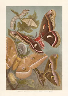
Silkmoth, chromolithograph, published in 1897Silkmoth: 1) Domestic silkmoth (Bombyx mori) with caterpillar, cocoons, and eggs; 2) Cecropia moth (Hyalophora cecropia, or Saturnia Cecropia); 3) Chinese Oak Silkmoth (Antheraea pernyi)
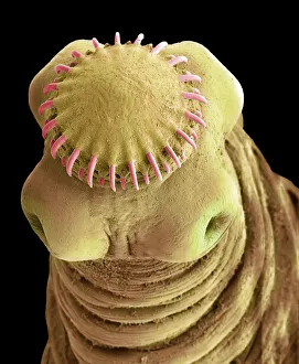
Tapeworm cysticercus, SEMTapeworm cysticercus. Scanning electron micrograph (SEM) of the head of the bladder worm or cysticercus stage of the pork tapeworm (Taenia solium), an intestinal parasite
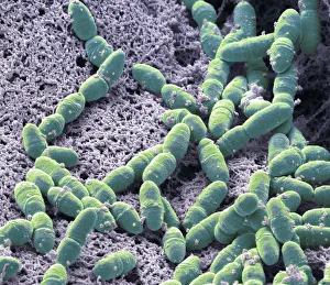
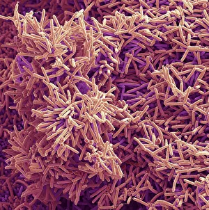
Streptococcus mutans, SEMStreptococcus mutans. Coloured scanning electron micrograph (SEM). S. mutans is a coccoid shaped, Gram-positive, anaerobic bacteria that is part of the normal bacteria flora of the mouth
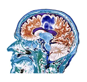
Glioblastoma brain cancer, CT scanGlioblastoma brain cancer. Coloured computed tomography (CT) scan of a section through the brain of an 84-year-old female patient with glioblastoma (blue, top)
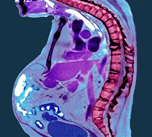
Spine in ankylosing spondylitis, X-raySpine in ankylosing spondylitis. Coloured X-ray of a section through the thoracic spine of a 74-year-old male patient with ankylosing spondylitis
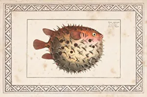
Birdbeak burrfish or Spotted Porcupinefish from 1797Birdbeak burrfish or prickly bottlefish, Cyclichthys orbicularis ( Orbicular diodon, Diodon orbicularis ) Spotted Porcupinefish
Plaque-forming bacteria, SEMPlaque-forming bacteria, coloured scanning electron micrograph (SEM). Plaque consists of a film of bacteria embedded in a glycoprotein matrix
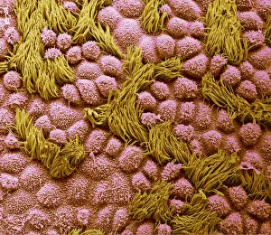
Fallopian tube, SEMFallopian tube. Coloured scanning electron micrograph (SEM) of the surface of a human fallopian tube. Fallopian tubes are ducts that lead from the ovaries to the uterus
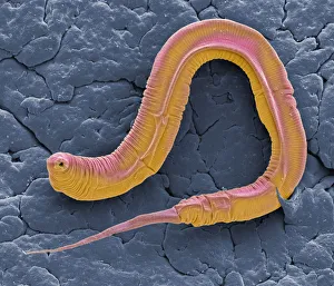
C elegans, SEMCaenorhabditis elegans worm, coloured scanning electron micrograph (SEM). C. elegans is a soil-dwelling hermaphrodite nematode worm and one of the most studied animals in biological
Handwriting of Hans Joachim von Zieten, general and confidante of Frederick the GreatIllustration from 19th century
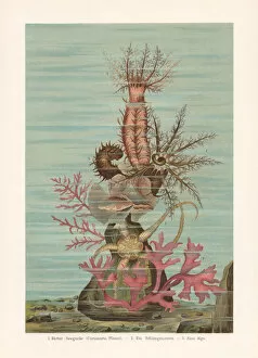
Sea ??cucumber, chromolithograph, published in 1897Sea ??cucumber: 1) Oloturia (Cucumaria planci); 2) Serpents table brittle star (Ophiura albida); 3) Seaweed. Chromolithograph, published in 1897
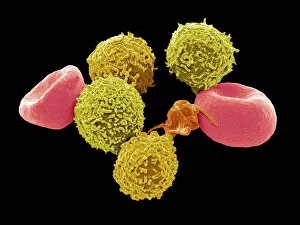
Blood cells, SEMBlood cells. Coloured scanning electron micrograph (SEM) of human red blood cells (erythrocytes, red), white blood cells (leukocytes, yellow), and platelets (thrombocyte, orange)