mail_outline sales@mediastorehouse.com
1,262 items
Digital illustration of male human brain
Digital cross section illustration of localization of source of sound in human brain
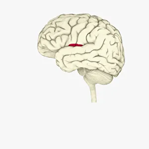
Digital illustration of insula in human brain highlighted in red
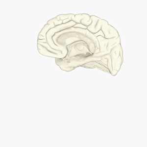
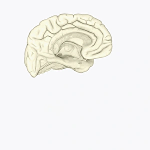
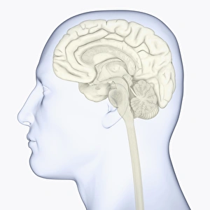
Digital illustration of human brain showing corpus collosum and cingulate gyrus on medial surface of human brain
Digital illustration of head in profile showing face recognition area and amygdala in brain
Digital illustration of head in profile showing lateral view of cortex in brain
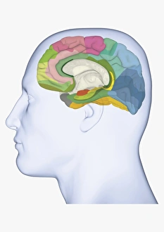
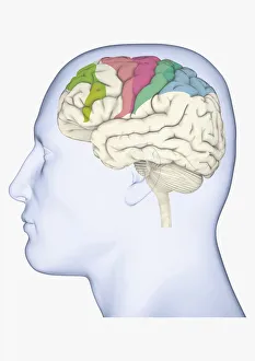
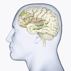
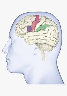
Digital illustration of head in profile showing medial Brodmann areas of human brain highlighted in different colours
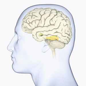
Digital illustration of head showing location of hippocampus in human brain
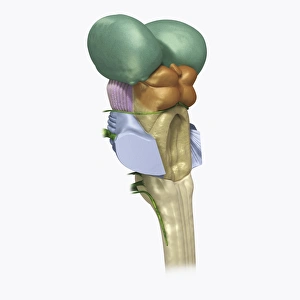
Digital illustration of human brain stem with cerebellum removed revealing medulla and axons
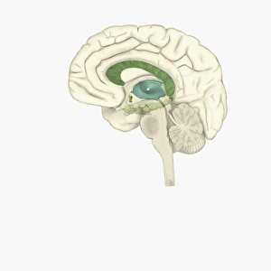
Digital illustration of head in profile showing brain highlighting thalamus in green and hypothalamus and pituitary gland in blue
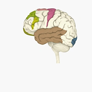
Digital illustration of head in profile showing brain with areas of movement highlighted in green, pink, blue and green
Digital illustration of human head in profile showing brain with frontal cortex highlighted in brown and supplementary motor area in pink
Digital illustration of head in profile showing motor cortex (pink), frontal area and amygdala (green), auditory cortex (orange), and visual cortex in brain
Digital illustration of head in profile showing thalamus (grey), and frontal lobe (green) in brain
Digital illustration of head in profile showing visual cortex (blue), motor cortex (pink), auditory cortex (orange), frontal area and amygdala (green) brain
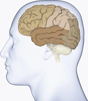
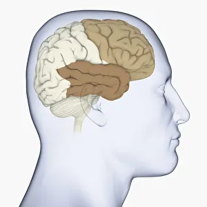
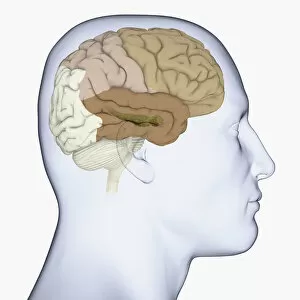
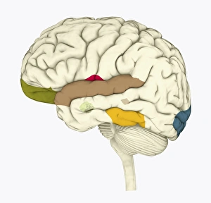
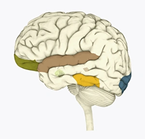
Digital illustration of head in profile showing frontal lobe and temporal lobe in brain
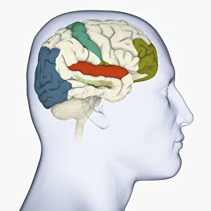
Digital illustration of head in profile showing frontal, parietal and temporal lobes, and hippocampus (green) in brain
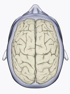
Digital illustration of head showing left and right areas of brain seen from above
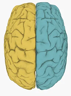
Digital illustration of left hemisphere (yellow) and right hemisphere (blue) of human brain
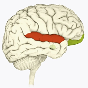
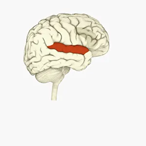
Digital illustration of superior temporal sulcas (red), orbitofrontal cortex and amygdala (green), in human brain
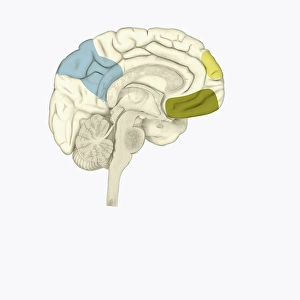
Digital illustration of posterior cingulate cortex (blue), medial frontal gyrus (yellow), and orbitofrontal prefrontal cortex (green) in human brain
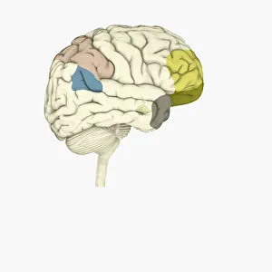
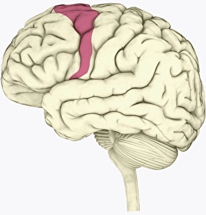
Digital illustration of parietal lobe (green), posterior superior temporal sulcas (blue), temperal pole (grey), dorsolateral prefrontal cortex, amygdala
Digital cross section illustration of human brain
Digital illustration of emotional response areas in human brain
Digital illustration of anterior cingulate cortex (grey), and medial frontal cortex (green) in human brain
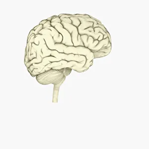
Digital illustration of human brain
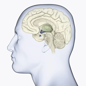
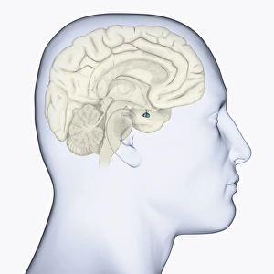
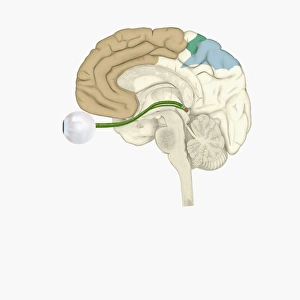
Digital illustration of head in profile showing pituitary gland in brain highlighted in blue
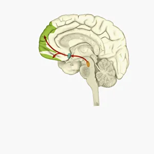
Digital illustration of reward pathway in human brain
Digital illustration of human brain associated with full awareness
Digital illustration of areas of information highlighted in human brain
Digital illustration of human brain with orbitofrontal cortex and amygdala highlighted in green
Digital illustration of head in profile showing areas of brain used for processing emotion
Digital illustration of head in profile showing brain
Digital illustration of head in profile showing mirror neurons in human brain highlighted in purple, pink and green
Digital illustration of head in profile showing area of mirror neuron in brain highlighted in orange
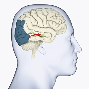
Digital illustration of head in profile showing direction of sensory signals from visual cortex to hippocampus in brain
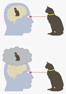
Digital illustration of monism and dualism in human brain
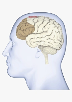
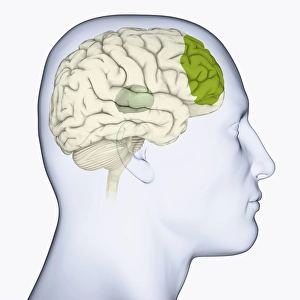
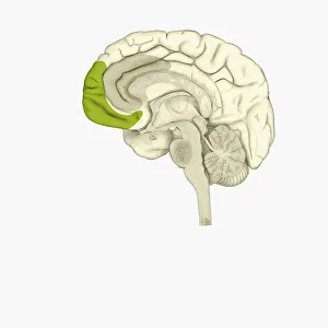
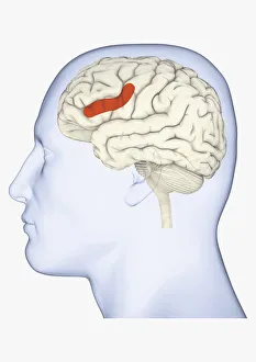
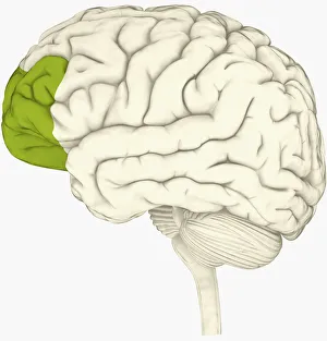
Digital illustration of prefrontal cortex of human brain highlighted in green
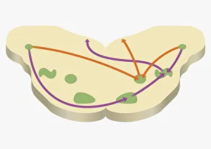
Digital illustration of various areas of cortex in human brain receiving input from sense organs
Digital illustration of crucial parts of highlighted in human brain
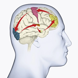
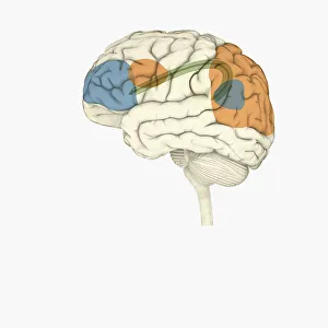
Digital illustration of frontal lobe and parietal lobe areas (orange) in left hemispheres (blue), and pathway of data from parietal lobe to frontal lobe (green) in human brain
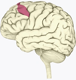
Digital illustration of lateral prefrontal cortex involved in decision making highlighted in green in human brain
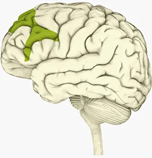
Digital illustration of premotor cortex involved in decision making highlighted in pink in human brain
Digital illustration of human head in profile showing brain
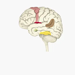
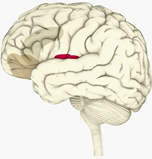
Digital illustration of right superior temporal sulcas, and anterior cingulate cortex highlighted in red and grey in human brain
Digital illustration of anterior insular, anterior cingulate cortex, and ventromedial prefrontal cortex highlighted in human brain
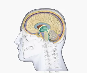
Digital illustration of head in profile showing skull, brain, and spine