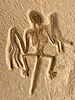
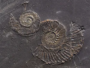
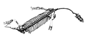
Fossil bat. Fossil Bute National Monument, Wyoming. USA
color image, photography, nature, nobody, vertical image, one animal, animal, wildlife, fauna, one, alone, individual, fossil, old, skeleton, bones, remains, ancient, imprint, bat
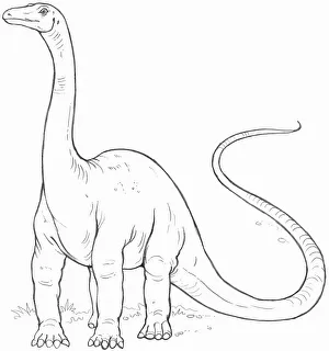
Line drawing of a diplodocus
Animal Themes, Black and White, Diplodocus, Full Length, Illustration and Painting, No People, One Animal, Palaeontology, Pencil Drawing, Side View, Square Image, Standing, Tail, White Background
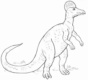
Line drawing of a Corythosaurus dinosaur
Animal Themes, Black and White, Corythosaurus, Full Length, Illustration and Painting, No People, One Animal, Palaeontology, Pencil Drawing, Side View, Square Image, Standing, White Background
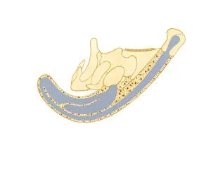
Illustration showing direction air passes through bony crest on top of Parasaurolophus skull
Air, Anatomy, Animal Bone, Animal Themes, Arrow Sign, Bony, Close-Up, Crest, Cross Section, Direction, Extinct, Illustration and Painting, No People, On Top Of, Paleontology, Parasaurolophus
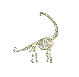
Illustration of skeleton of Brachiosaurus dinosaur
Anatomy, Animal Skeleton, Animal Themes, Brachiosaurus, Dinosaur, Extinct, Full Length, Herbivorous, Illustration and Painting, Jurassic, Long, Looking Away, Mouth Open, Neck, No People, One Animal
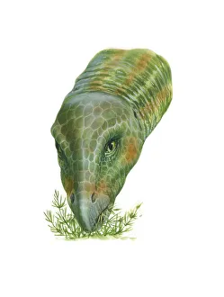
Illustration of Hypsilophodon dinosaur feeding on plants
Animal Behaviour, Animal Head, Animal Themes, Beak, Camouflage, Close-Up, Cretaceous, Extinct, Feeding, Food Chain, Front View, Green, Head and Shoulders, Herbivorous, Hypsilophodon
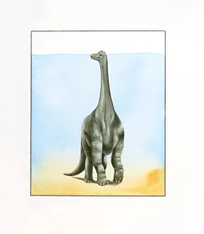
Illustration of large Barosaurus dinosaur walking underwater with top of head peeking above surface on long neck
Above, Animal Behaviour, Animal Head, Animal Themes, Barosaurus, Dinosaur, Extinct, Front View, Full Length, Herbivorous, Illustration and Painting, Jurassic, Large, Long, Motion, Neck, No People