mail_outline sales@mediastorehouse.com
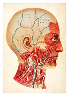
Human Anatomy illustration 1891The Practical Physician
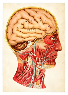
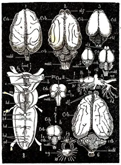
Human Brain illustration 1891The Practical Physician
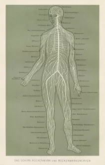
Nerves anatomy engraving 1857Rank, johannes - The human being. 1 - 1894
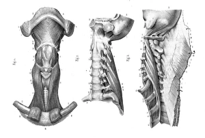
Neck vertebra anatomy engraving 1866Atlas d anatomie descriptive du corps humain C. Bonamy - Paul Broca Victor Masson et Fils Paris 1866
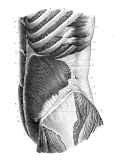
Frontal trunk anatomy engraving 1866Atlas d anatomie descriptive du corps humain C. Bonamy - Paul Broca Victor Masson et Fils Paris 1866
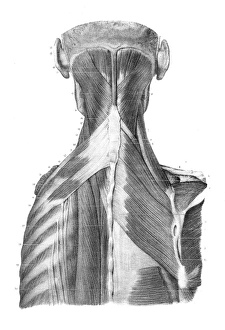
Back trunk anatomy engraving 1866Atlas d anatomie descriptive du corps humain C. Bonamy - Paul Broca Victor Masson et Fils Paris 1866
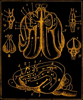
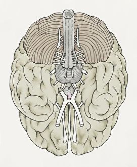
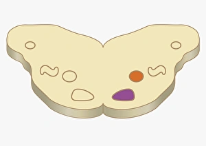
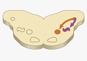
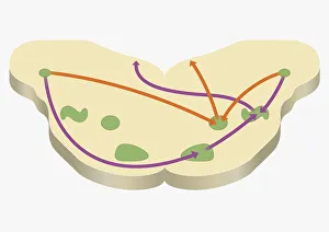
CNS partsMiddle and anterior-posterior section of the brain
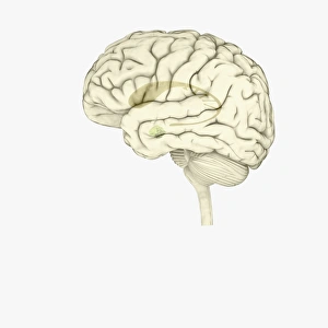
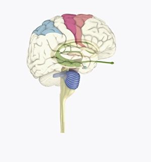
Brain anatomyIllustration of a brain anatomy
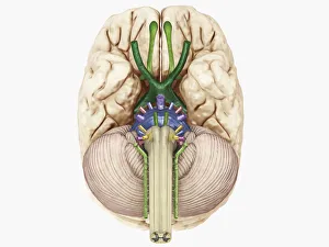
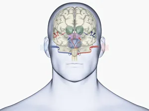
Illustration of cranial nerves of human brain
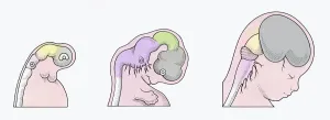
Illustration of three stages of Neurogenesis responsible for populating growth of brain in human embryo
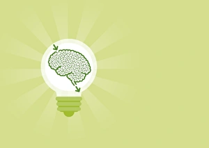
Digital illustration of human brain inside lightbulb
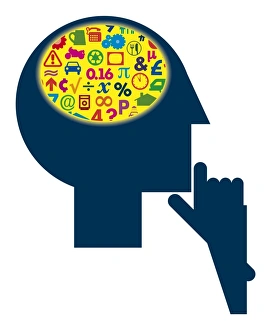
Digital illustration of signs and symbols inside head representing different areas of the human brain
Illustration showing connection of thyroid gland and pituitary gland to brain of pre-adolescent girl
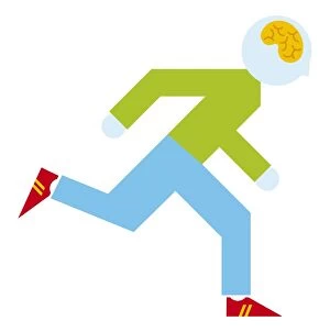
Digital illustration of running man showing brain inside head
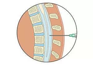
Digital illustration of lumbar puncture using spinal needle inserted into lumbar vertebrae and dura mater
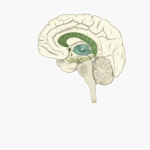
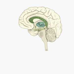
Digital illustration of striatum and amygdala highlighted in human brain
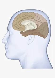
Digital illustration of head in profile showing medial view of cortex in human brain
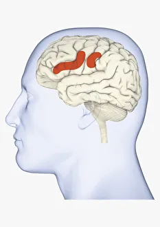
Digital illustration of head in profile showing areas of mirror neuron in brain highlighted in orange
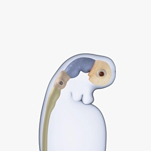
Digital illustration of neural tube, rudimentary eye and ear buds of three week old human embryo
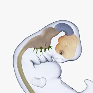
Digital illustration of 7 week old human embryo
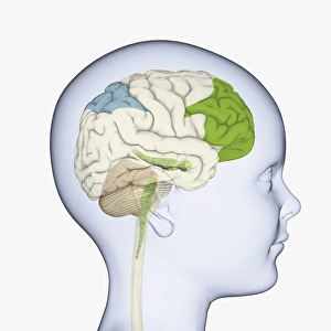
Digital illustration of childs head in profile highlighting parts of brain
Digital illustration of same side processing of smell from nostril to human brain
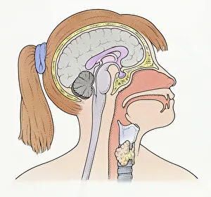
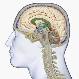
Digital illustration of head in profile showing cross section of brain, neck vertebra and spine
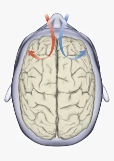
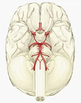
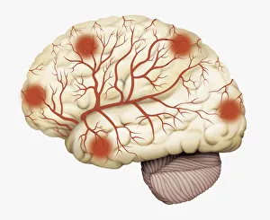
Digital illustration showing arteries in human brain
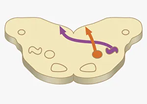
Digital cross section illustration of impulses from lower brain stem passing to inferior colliculus of midbrain
Digital illustration of cranial nerves linked to human brain
Cross section digital illustration of human brain showing caudate nucleus, putamen, external globus pallidus, and internal globus pallidus
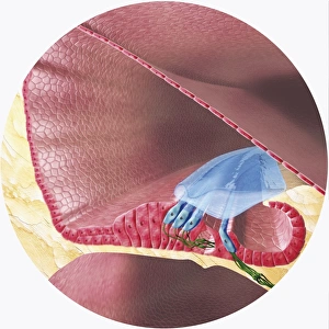
Digital illustration of Corti organ found in cochlea of human ear
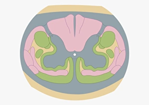
Cross section digital illustration of spinal nerve fibres and convey motor signals highlighted in pink and green
Digital illustration of human brain showing blood vessels, and areas of dead tissue highlighted in red
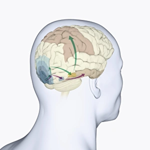
Digital illustration of head in profile showing dorsal and ventral pathways of brain
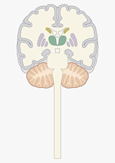
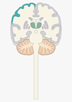
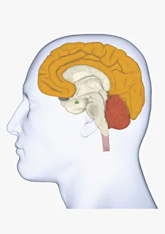
Cross section digital illustration of brain highlighting cerebral cortex, thalamus, and brain stem
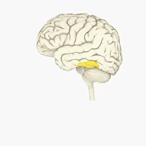
Digital illustration of human brain showing face-recognition area highlighted in yellow
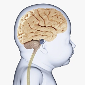
Digital illustration of head of baby in profile showing brain
Digital illustration head in profile with cerebrum and cortex highlighted
Digital illustration of human brain with sound entering via brain stem and thalamus to auditory cortex
Digital cross section illustration of medial nucleus and lateral superior olive in human brain
Digital illustration of woman with head in profile showing brain connected to spinal cord
Digital cross section illustration of impulses passing from nearer cochlea nucleus to lateral superior olive
Digital cross section illustration of area signal received in cell of ventral cochlea in human brain
Digital illustration of highlighted areas in human brain affected by motor disorders
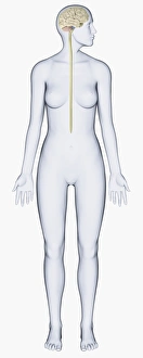
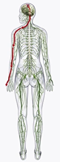
Digital illustration of of human nervous system
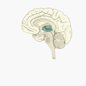
Digital illustration of thalamus in human brain highlighted in green
Digital illustration of female human brain
Digital illustration of male human brain
Digital cross section illustration of localization of source of sound in human brain